X-ray imaging device

An imaging device and X-ray technology, which can be used in the control of radiological diagnosis equipment, transmission of radiological diagnosis data, instruments used for radiological diagnosis, etc., and can solve problems such as image blurring.

Inactive Publication Date: 2020-04-10
TOSHIBA IT & CONTROL SYSTEMS CORP
View PDF6 Cites 0 Cited by
  • Summary
  • Abstract
  • Description
  • Claims
  • Application Information

AI Technical Summary

Problems solved by technology

However, in the prior art, such a problem cannot be solved, and the operator has to determine the appropriate tube voltage value v and tube current value i while judging the blurring of the image by visual inspection.

Method used


Image

  • X-ray imaging device
  • X-ray imaging device
  • X-ray imaging device

Examples


no. 2 Embodiment approach

[0147] In the second embodiment, in addition to the processing of the first embodiment, a mechanical processing of changing the position of each part of the imaging device, that is, a hardware processing (hard) processing is added to the captured image, thereby obtaining a medium resolution or a SN ratio. Either of the enhanced images. Other configurations of the second embodiment are the same as those of the first embodiment. The same reference numerals are assigned to the same configuration as in the first embodiment, and description thereof will be omitted.

[0148] Such as Figure 4 As shown in the flowchart of FIG. 2 , in the second embodiment, it is selected whether to perform resolution or SN ratio correction processing on the image captured in the first embodiment ( S51 ). The selection is performed by the operator outputting a predetermined command from the input device 13 to the mechanism control unit 11 . If the correcting process is selected (Yes (YES) in S51), ...

no. 3 Embodiment approach

[0153] In the third embodiment, instead of the resolution correction or SN ratio correction processing in the second embodiment, the image processing unit 9f performs software-based image processing such as contour enhancement or noise removal on the captured image, thereby obtaining improved visibility. Image. Other configurations of the third embodiment are the same as those of the second embodiment. The same reference numerals are assigned to the same configuration as in the second embodiment, and description thereof will be omitted.

[0154] Such as Figure 5As shown in the flow chart of , in the third embodiment, it is selected whether to perform correction processing of edge enhancement or noise removal on the image captured in the first embodiment (S53). The selection is performed by the operator outputting a predetermined command from the input device 13 to the mechanism control unit 11 . If the correction process is selected (Yes in S53), the mechanism control unit...


PUM


Abstract

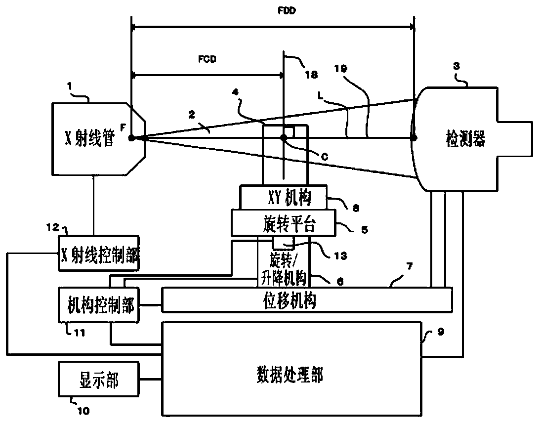
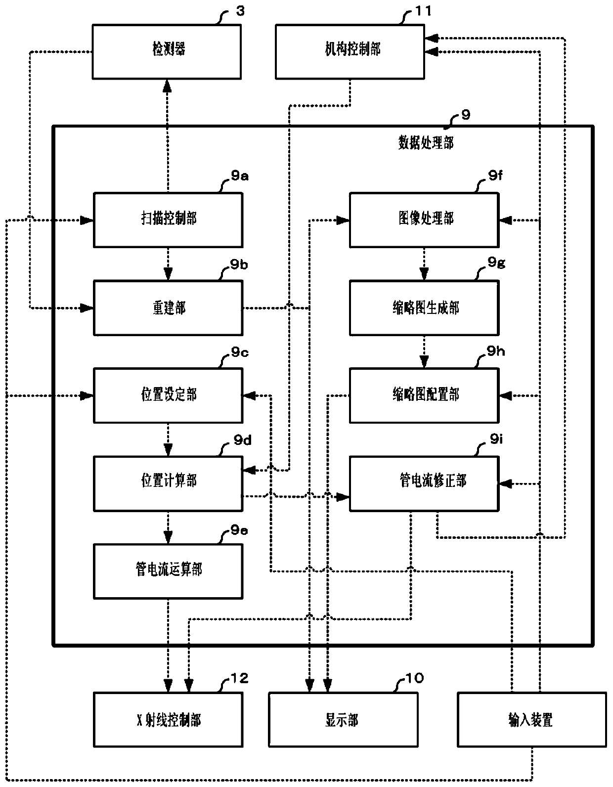
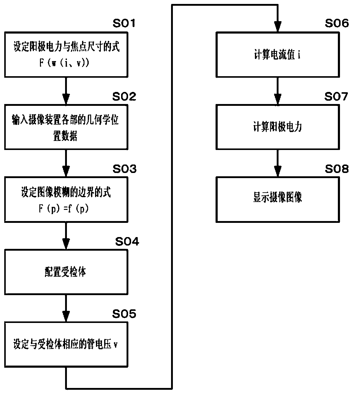
Provided is an X-ray imaging device that automatically adjusts a tube current value in accordance with changes in the one pixel size and the tube voltage value v of an acquired image. A position setting unit (9c) sets the geometric positional relationship among the X-ray generator (1), the subject (4), and the X-ray detector (3). A tube current calculation unit (9e) obtains change characteristicsbetween one pixel size of an image and a focal point size with respect to anode power of an X-ray generator (1) based on the geometric position of the X-ray generator (1), the subject (4), and the X-ray detector (3), and calculates the tube current value i on the basis of a tube voltage value v determined from the subject (4). The X-ray control unit (12) determines the anode power of the X-ray generator (1) on the basis of the tube current value i and the tube voltage value v, and controls the output of the X-ray generator (1) on the basis of the determined anode power.

Description

technical field [0001] Embodiments of the present invention relate to an X-ray imaging apparatus that automatically sets radiation conditions based on the size of one pixel of an acquired image. Background technique [0002] In an X-ray imaging device such as an X-ray fluoroscopy device or an X-ray computed tomography (CT) device, for example, as shown in Patent Document 1 to Patent Document 3, the tube voltage value v and the tube current value i are used. Two parameters to control the X-ray projection conditions. When most X-ray generators increase the anode power (tube voltage value v×tube current value i), the focus size of X-rays will become larger, resulting in blurred images. [0003] In the conventional device, the operator must input the values ​​of tube voltage and tube current into the device to adjust the focus size while observing the blurring of the image. At this time, if the focus size is to be kept small to avoid blurring of the image, the anode power will...

Claims


Application Information

Patent Timeline
Patent Type & Authority Applications(China)
IPC IPC(8): G01N23/04
CPCG01N23/04A61B6/54A61B6/56A61B6/5205A61B6/4429
Inventor 原拓生长野雅実
Owner TOSHIBA IT & CONTROL SYSTEMS CORP
Why Eureka
Try Eureka