
Medical image processing method, medical image processing device, storage medium and computer equipment

A medical image and processing method technology, applied in the field of image processing, can solve the problems of missed detection of image processing results, unfavorable and accurate diagnosis, and inability to obtain detection data.

Pending Publication Date: 2020-05-08
SHANGHAI UNITED IMAGING INTELLIGENT MEDICAL TECH CO LTD
View PDF6 Cites 4 Cited by
  • Summary
  • Abstract
  • Description
  • Claims
  • Application Information

AI Technical Summary

Problems solved by technology

[0003] In the prior art, when automatically processing medical images, the model used is usually a single-disease model, that is, it can only detect a single disease condition. However, the image processing results obtained by the existing medical image processing models have gaps Therefore, reliable test data cannot be obtained to support doctors in further diagnosis, which is not conducive to accurate diagnosis in the later stage

Method used


Image

  • Medical image processing method, medical image processing device, storage medium and computer equipment
  • Medical image processing method, medical image processing device, storage medium and computer equipment
  • Medical image processing method, medical image processing device, storage medium and computer equipment

Examples


Embodiment Construction

[0030] In order to make the purpose, technical solution and advantages of the present application clearer, the present application will be further described in detail below in conjunction with the accompanying drawings and embodiments. It should be understood that the specific embodiments described here are only used to explain the present application, and are not intended to limit the present application.

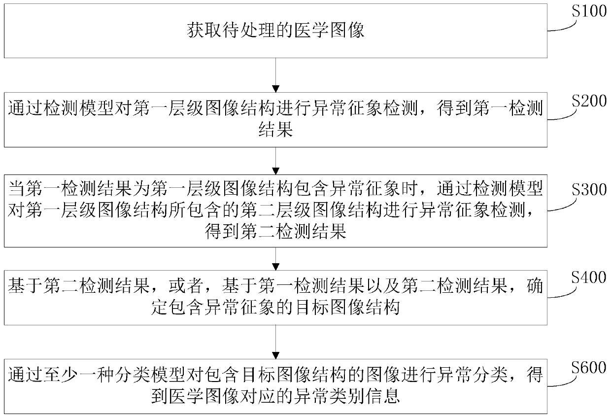
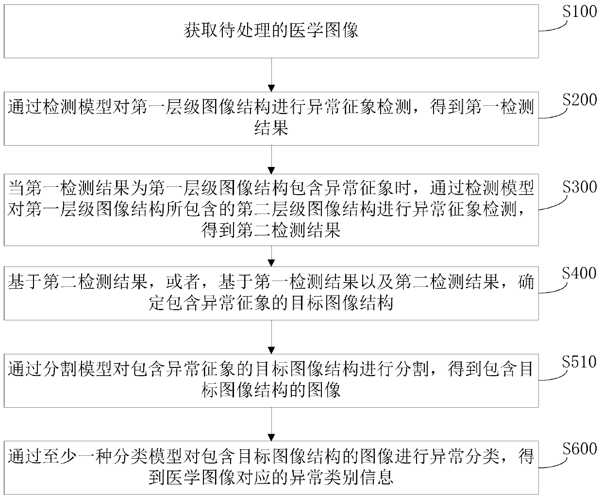
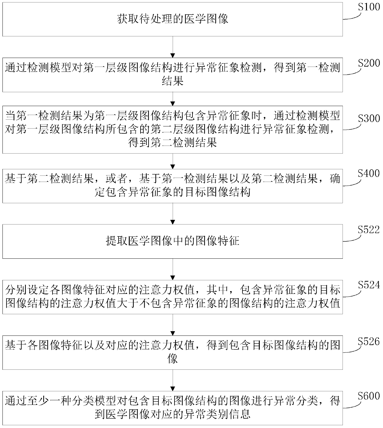
[0031] In one embodiment, such as figure 1 As shown, a medical image processing method is provided, and the method is applied to a processor capable of medical image processing as an example for explanation. The method mainly includes the following steps:

[0032] Step S100, acquiring medical images to be processed.

[0033] Wherein, the medical image includes image structures of different levels, and different levels specifically refer to different ranges and levels of the image structures. For example, a medical image includes at least a first-level image structure and...


PUM


Abstract

The invention relates to a medical image processing method, a medical image processing device, a storage medium and computer equipment. According to the invention, during processing of a medical image, when it is detected that a first-level image structure contains abnormal signs, abnormal sign detection is continuously conducted on the second-level image structure contained in the first-level image structure, that is, abnormal sign detection is conducted on all the image structures according to the sequence from the high level to the low level, so that the abnormal detection can be comprehensive; and the target image structure containing the abnormal signs is determined based on the detection result, and then abnormal classification is performed through at least one classification model,so that the accuracy of the abnormal classification result can be enhanced, reliable abnormal category information can be obtained to support the doctor to perform further diagnosis, and the doctor can perform accurate diagnosis in the later period.

Description

technical field [0001] The present application relates to the technical field of image processing, in particular to a medical image processing method, device, storage medium and computer equipment. Background technique [0002] With the rapid development of medical imaging technology, various medical scanning images have been produced, such as PET (Positron Emission Computed Tomography, Positron Emission Computed Tomography) images, CT (Computed Tomography, electronic computer tomography) images, MRI (Magnetic Resonance Imaging, magnetic resonance imaging) images, etc. Medical images are an important reference for doctors to diagnose. Therefore, in order to improve the efficiency of diagnosis, the automatic processing of medical images is of great significance. [0003] In the prior art, when automatically processing medical images, the model used is usually a single-disease model, that is, it can only detect a single disease condition. However, the image processing results...

Claims


Application Information

Patent Timeline
IPC IPC(8): G16H30/20G16H30/40G06T7/00G06T7/11G06T11/00G06K9/62
CPCG16H30/20G16H30/40G06T7/0012G06T7/11G06T11/005G06T2207/30004G06F18/241
Inventor 薛忠曹晓欢
Owner SHANGHAI UNITED IMAGING INTELLIGENT MEDICAL TECH CO LTD
Learn More