
Eureka
For R&D, Eureka makes reading and utilizing patents & technical documents easy.
Eureka AIR
Designed for self-driven R&D workflows. Generate viable solutions, solve complex R&D challenges, empower your innovation with AI.

Eureka Materials
Designed for material experts only. Revolutionize your material R&D, from search, analyze, to developing new materials.

TechResearch
Generate reliable direction feasibility study reports for your R&D in just a few steps.

TechSeek
Discover and master advanced knowledge NOW. Basics, ideas, possibilities, all at once.

TechMind
As an expert in R&D Theories, TechMind can generates customized viable solutions instantly.

TechRisk
Analyze your overall solution with one click, know your potential R&D risks in advance.

TechMonitor
Get weekly tech updates, stay abreast of the latest tech innovations and key insights.
Method and device for determining coronary artery branch where calcified area is located, server and medium
A determination method and technique of coronary artery, applied in the medical field, can solve the problem of indistinguishable muscle tissue, etc., and achieve the effect of accurate determination
- Summary
- Abstract
- Description
- Claims
- Application Information
AI Technical Summary
Problems solved by technology
Method used
Image

Examples
Embodiment 1
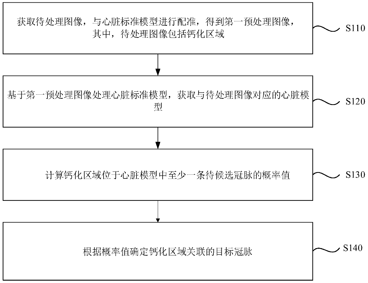
[0029] figure 1 It is a schematic flowchart of a method for determining the coronary artery branch where the calcified area is located in Embodiment 1 of the present invention. This embodiment can be applied to quickly determine the coronary artery branch where the calcified area is located. The device for determining the branch is executed, and the device can be implemented in the form of software and / or hardware.
[0030] like figure 1 Described, the method of the present embodiment comprises:
[0031] S110. Acquire the image to be processed, and perform registration with the heart standard model to obtain a first pre-processed image, wherein the image to be processed includes a calcification area.
[0032] In order to clearly introduce the technical solution of this embodiment, the part to be scanned is the heart as an example.
[0033] Wherein, the original image obtained after scanning the part to be scanned is used as the image to be processed. The scanned part may b...
Embodiment 2
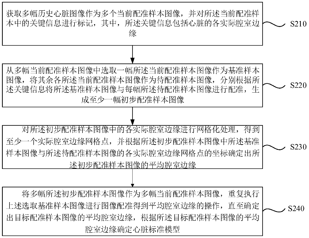
[0054] Before determining which branch of the coronary artery the calcified area in the image to be processed is located in, it is also necessary to determine the standard model of the heart and the chamber classifier corresponding to each chamber, so as to register the image to be processed based on the standard heart model, A heart model is obtained to determine the target coronary artery to which the calcified region belongs according to the heart model. figure 2 It is a schematic flowchart of a method for determining the coronary artery branch where the calcified area is provided in the second embodiment of the present invention.
[0055] like figure 2 As shown, the method includes:
[0056] S210. Acquire multiple historical heart images as multiple current registration sample images, and mark key information in the current registration samples, where the key information includes actual chamber edges of the heart.
[0057] Wherein, the plurality of historical cardiac i...
Embodiment 3
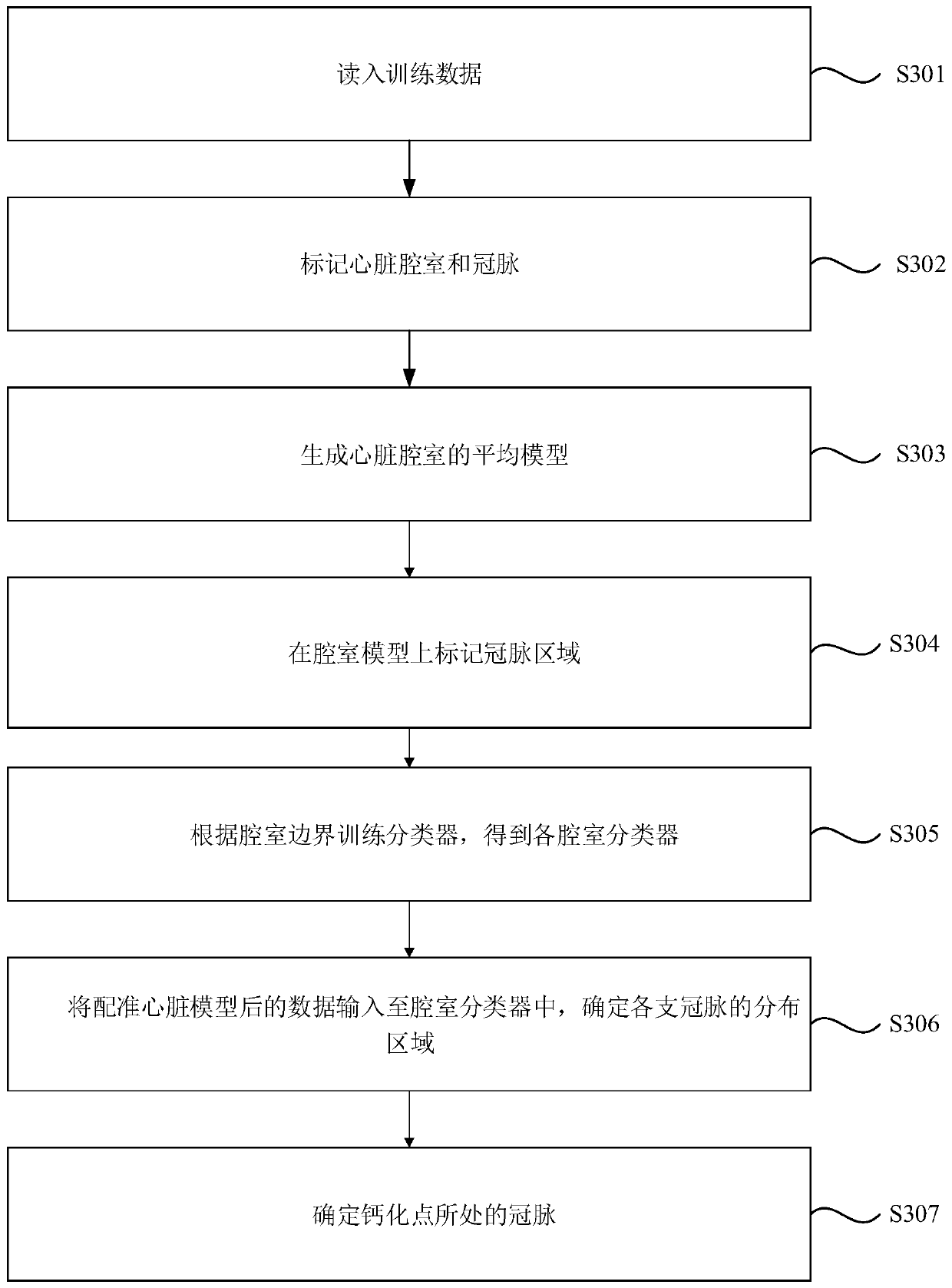
[0075] As a preferred embodiment of the above embodiment, image 3 It is a schematic flowchart of a method for determining the coronary artery branch where the calcified region is provided by the embodiment of the present invention.
[0076] like image 3 Described, the method of the present embodiment comprises:
[0077] S301. Reading in training data.
[0078] Select a certain amount of training data, optional, thousands or tens of thousands, etc. The training data can be understood as heart scan images.
[0079] It should be noted that in order to obtain the heart model, the size and resolution of the heart image need to be consistent.
[0080] Specifically, a preset number of training sample images are acquired to train the cardiac standard model.
[0081] S302 , marking heart chambers and coronary arteries.
[0082]Before training the data, the left ventricle, right ventricle, left atrium, right atrium, pulmonary artery root, aortic base, etc. structures of the hear...
PUM
 Abstract
Description
Claims
Application Information
 - R&D Engineer
- R&D Manager
- IP Professional
- Industry Leading Data Capabilities
- Powerful AI technology
- Patent DNA Extraction
Browse by: Latest US Patents, China's latest patents, Technical Efficacy Thesaurus, Application Domain, Technology Topic, Popular Technical Reports.
© 2024 PatSnap. All rights reserved.Legal|Privacy policy|Modern Slavery Act Transparency Statement|Sitemap|About US| Contact US: help@patsnap.com