Liver cancer image feature extraction and pathological classification method and device based on imaging omics

A technology of image feature extraction and radiomics, applied in the field of medical image processing, can solve the problem of rough evaluation of liver cancer differentiation

Active Publication Date: 2020-06-05
ZHEJIANG UNIV
View PDF0 Cites 14 Cited by
  • Summary
  • Abstract
  • Description
  • Claims
  • Application Information

AI Technical Summary

Problems solved by technology

Previous studies have shown that radiomics features derived from computed tomography (CT) magnetic resonance imaging (MRI) are helpful for the identification of pathological gra

Method used


Image

  • Liver cancer image feature extraction and pathological classification method and device based on imaging omics
  • Liver cancer image feature extraction and pathological classification method and device based on imaging omics
  • Liver cancer image feature extraction and pathological classification method and device based on imaging omics

Examples


Embodiment Construction

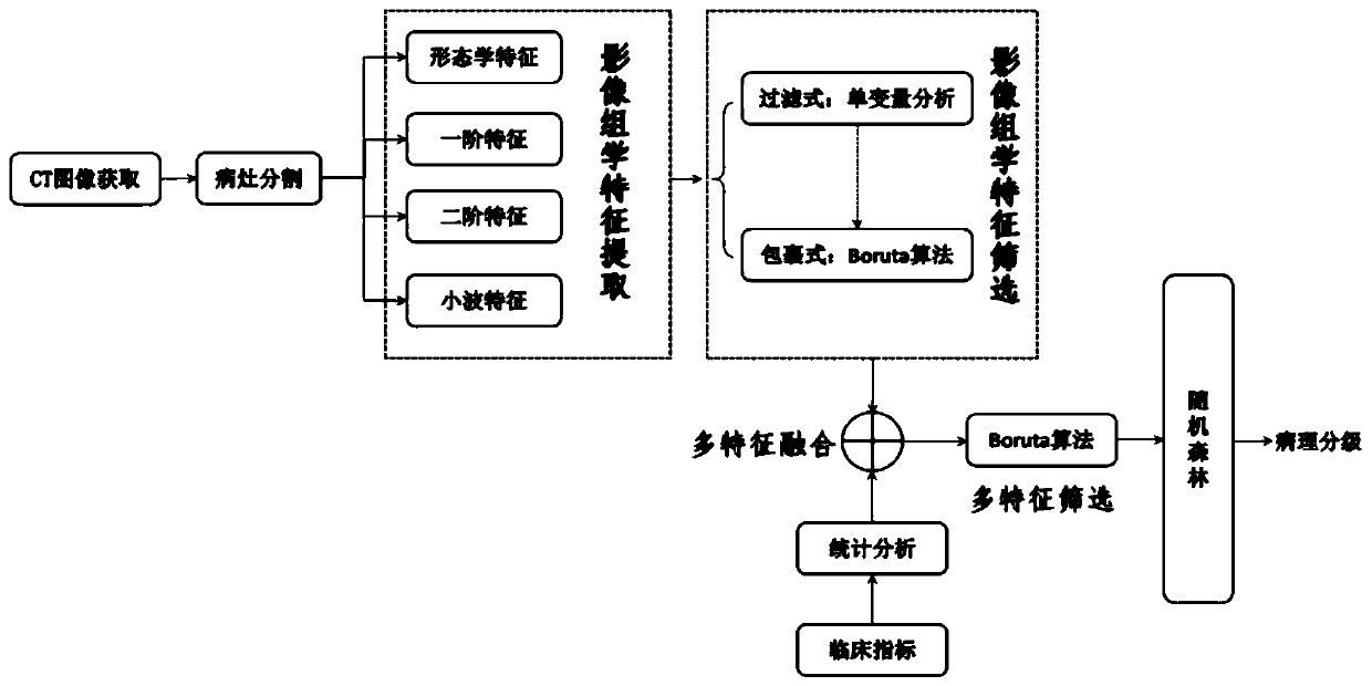
[0094] The method of the present invention will be further described below in conjunction with the accompanying drawings.

[0095] Step (1). Liver cancer images and their corresponding pathological classification labels are used as training data sets;

[0096] Step (2). Using the GrowCut algorithm to achieve semi-automatic segmentation of the liver cancer lesion area. in grid position (pixels or voxels in image processing). The cellular automaton is expressed as a triple A=(S,N,δ), where A represents a cellular automaton model, S is a non-empty state set, N is the domain system, δ:S N → S is the local transition function, which defines the rules for computing the state of a cell at t+1 time steps given the state of a neighborhood cell at time step t. The neighborhood system N used is the von Neumann neighborhood:

[0097]

[0098] cell state where l p Indicates the label of the current cell, θ p is the current cell strength, is the feature vector of the current c...


PUM


Abstract

The invention discloses a liver cancer image feature extraction and pathological classification method and device based on imaging omics. The method comprises the following steps: 1) collecting a patient clinical image meeting the standard, and sketching a liver cancer lesion area of the collected image by adopting a Growcut semi-automatic segmentation method; 2) performing different levels of image omics feature extraction on the segmented lesion area; 3) feature screening: starting from a filtering method, extracting non-redundant features strongly related to the classification targets by adopting a filtering Boruta algorithm; 4) in combination with clinical indexes of the patient, filtering out significant and undifferentiated features through preliminary statistical analysis, and thenfusing the image omics features to perform next Boruta screening; and 5) training on a random forest by using the finally screened features to obtain classification labels, and completing prediction of pathological classification of liver cancer. Compared with a clinically traditional biopsy method, the method provided by the invention has the characteristics of non-invasion, safety and stability,and is expected to become an effective preoperative evaluation tool for clinic.

Description

technical field [0001] The invention belongs to the technical field of medical image processing, and in particular relates to a radiomics-based liver cancer image feature extraction and pathological classification method. Background technique [0002] Liver cancer is one of the leading causes of cancer death in the world, ranking the 7th and 3rd in the global tumor incidence and mortality, respectively. China accounts for 50% of the world's new liver cancer cases, endangering national health and causing heavy economic burden to families and society. Individualized comprehensive treatment according to the different stages of liver cancer is the key to improving the curative effect. At present, the traditional influencing factors for judging the staging of liver cancer include tumor size, number, and depth of tumor invasion, etc., and cannot reflect the intrinsic heterogeneity of the tumor, which determines the clinical biological behavior and prognosis of the tumor. The pat...

Claims


Application Information

Patent Timeline
IPC IPC(8): G06K9/62G06T7/00G06T7/11
CPCG06T7/11G06T7/0012G06T2207/30096G06T2207/30056G06F18/2113G06F18/24323G06F18/253G06F18/214
Inventor 丁勇阮世健邵嘉源丁越雷
Owner ZHEJIANG UNIV
Why Eureka
Try Eureka