Focus detection method, device and equipment, and storage medium

A detection method and lesion technology, applied in the field of lesion detection, can solve the problems of not considering three-dimensional context information and inaccurate measurement results.

Pending Publication Date: 2020-06-16
BEIJING SENSETIME TECH DEV CO LTD
View PDF3 Cites 2 Cited by
  • Summary
  • Abstract
  • Description
  • Claims
  • Application Information

AI Technical Summary

Problems solved by technology

However, most of the current lesion detection methods tend to only focus on the detection of certain lesion types, such as lung nodules, skin lesions, liver tumors, enlarged lymph nodes, colon polyps, etc. In addition, in the prior art, the measurement of lesions The judgment usually does not take into account the three-dimensional context information, resulting in inaccurate measurement results

Method used


Image

  • Focus detection method, device and equipment, and storage medium
  • Focus detection method, device and equipment, and storage medium
  • Focus detection method, device and equipment, and storage medium

Examples


Embodiment Construction

[0119] The following will clearly and completely describe the technical solutions in this application with reference to the accompanying drawings in this application. Obviously, the described embodiments are some of the embodiments of this application, but not all of them. Based on the embodiments in this application, all other embodiments obtained by persons of ordinary skill in the art without making creative efforts belong to the scope of protection of this application.

[0120] It should be understood that when used in this specification and the appended claims, the terms "comprising" and "comprises" indicate the presence of described features, integers, steps, operations, elements and / or components, but do not exclude one or Presence or addition of multiple other features, integers, steps, operations, elements, components and / or collections thereof.

[0121] It should also be understood that the terminology used in the specification of this application is for the purpose ...


PUM


Abstract

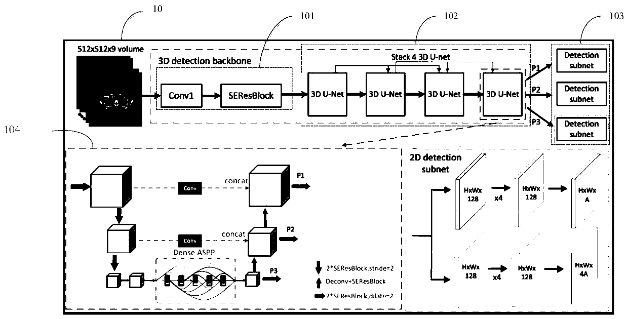
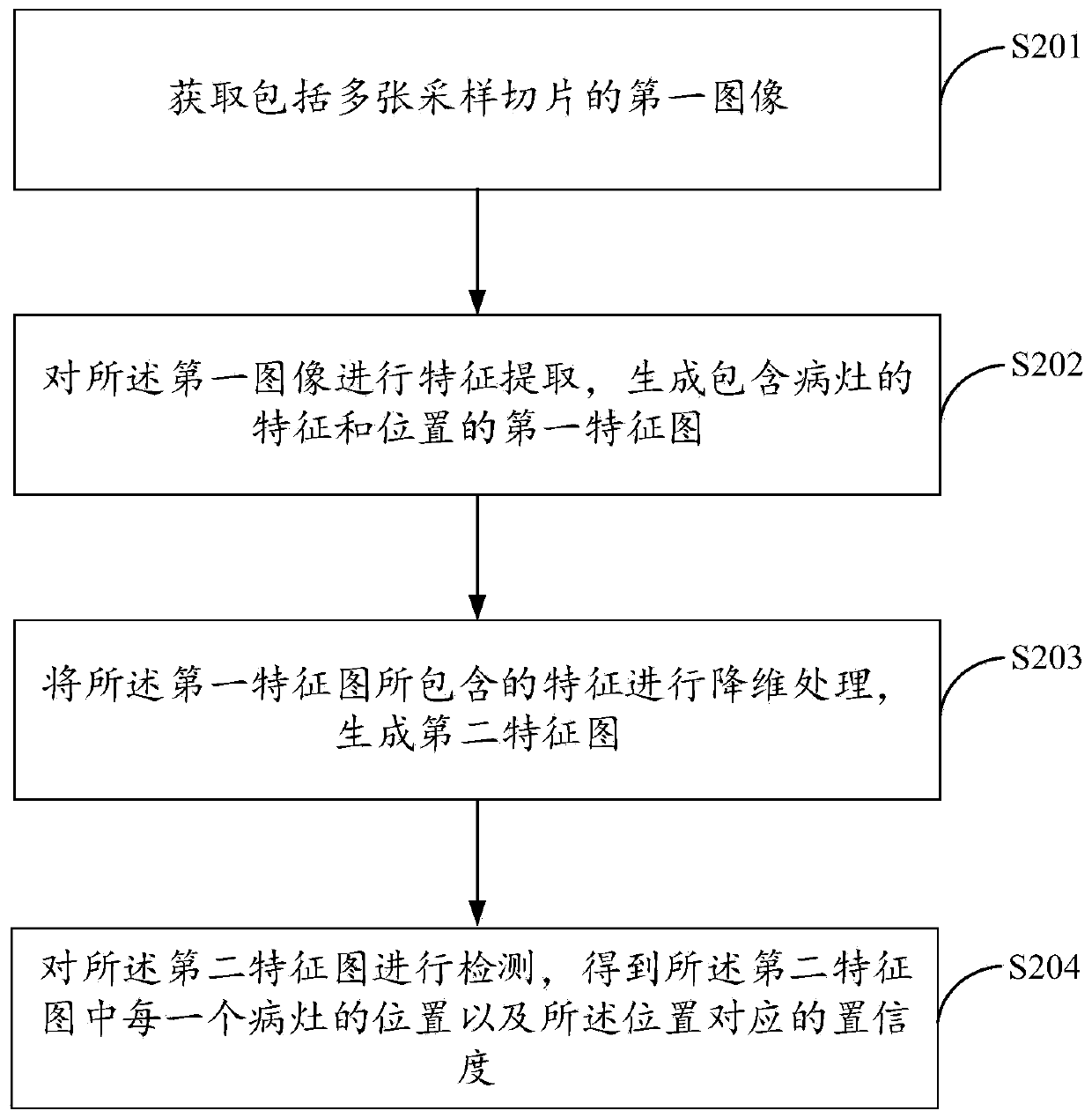
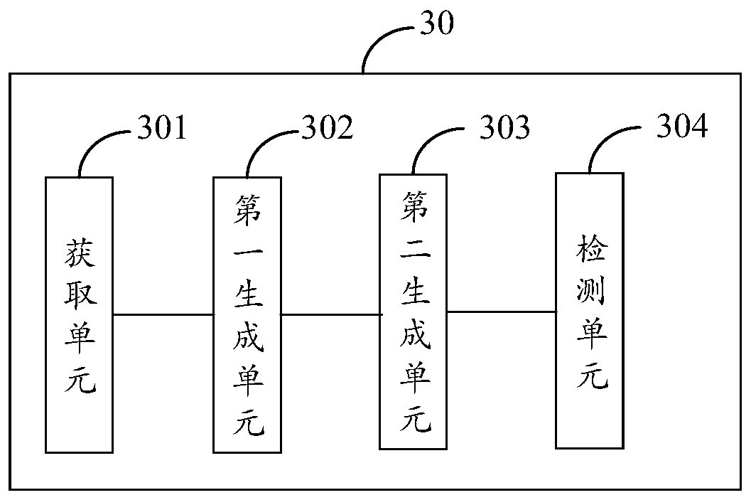
The invention discloses a focus detection method, device and equipment and a storage medium, and the method comprises the steps: obtaining a first image comprising a plurality of sampling slices, wherein the first image is a three-dimensional image comprising an X-axis dimension, a Y-axis dimension and a Z-axis dimension; performing feature extraction on the first image to generate a first featuremap containing features and positions of lesions; wherein the first feature map comprises three-dimensional features of an X-axis dimension, a Y-axis dimension and a Z-axis dimension; performing dimension reduction processing on features contained in the first feature map to generate a second feature map; wherein the second feature map comprises two-dimensional features of an X-axis dimension anda Y-axis dimension; and detecting the features of the second feature map to obtain the position of each lesion in the second feature map and the confidence coefficient corresponding to the position.By adopting the method and the device, the focus conditions of a plurality of parts in the body of the patient can be accurately detected, and primary cancer assessment of the whole body range of thepatient is realized.

Description

[0001] This application is a divisional application of a Chinese patent application with the application number 201811500631.4, the application date is December 7, 2018, and the invention name is "a lesion detection method, device and equipment". technical field [0002] The present application relates to the field of computer technology, and in particular to a method, device, equipment and storage medium for lesion detection. Background technique [0003] Computer aided diagnosis (CAD) refers to the automatic detection of lesions in re-imaging through imaging, medical image analysis technology and other possible physiological, biochemical and other means, combined with computer analysis and calculation. Practice has proved that computer-aided diagnosis has played a great positive role in improving the accuracy of diagnosis, reducing missed diagnosis and improving the work efficiency of doctors. Among them, the lesion refers to the part of the tissue or organ that is affecte...

Claims


Application Information

Patent Timeline
Patent Type & Authority Applications(China)
IPC IPC(8): G06T7/00G06T7/73G16H30/20G16H50/20
CPCG06T7/0012G06T7/73G16H30/20G16H50/20G06T2207/10081G06T2207/30004G06T2207/20081G06T2207/20084G16H30/40A61B6/032A61B6/5205G06T2207/30061G06T2207/30096G06T1/20G06N3/045G06T7/11G06N3/08G16H15/00A61B6/463A61B6/5217A61B6/5223G06T3/4046
Inventor 黄锐高云河
Owner BEIJING SENSETIME TECH DEV CO LTD
Why Eureka
Try Eureka