Medical image segmentation method and system based on deep learning, terminal and storage medium

A technology of medical imaging and deep learning, which is applied in the field of medical imaging and computer aids, can solve the problems of not being universal, not being used, and imaging, etc., and achieve the goal of improving accuracy and reliability, measurement efficiency, and diagnosis rate Effect

Pending Publication Date: 2020-07-10
BEIJING SHENRUI BOLIAN TECH CO LTD +1
View PDF7 Cites 7 Cited by
  • Summary
  • Abstract
  • Description
  • Claims
  • Application Information

AI Technical Summary

Problems solved by technology

However, these methods based on artificially designed features or thresholds are usually not very universal
For different devices, tube current tube voltage, using different scanning doses, and using different window widths and levels will all cause huge images on the segmentation results

Method used


Image

  • Medical image segmentation method and system based on deep learning, terminal and storage medium
  • Medical image segmentation method and system based on deep learning, terminal and storage medium
  • Medical image segmentation method and system based on deep learning, terminal and storage medium

Examples


Embodiment Construction

[0084] In order to make the purposes, technical solutions and advantages of the embodiments of the present application clearer, the technical solutions in the embodiments of the present application will be clearly and completely described below in conjunction with the drawings in the embodiments of the present application. Obviously, the described embodiments It is a part of the embodiments of this application, not all of them. Based on the embodiments in this application, all other embodiments obtained by persons of ordinary skill in the art without making creative efforts belong to the scope of protection of this application.

[0085] Please refer to figure 1 , figure 1 A flow chart of medical image segmentation based on deep learning provided by the embodiment of the present application, the method includes:

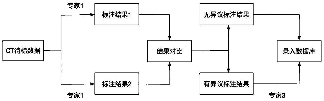
[0086] S101: Collect medical image data and perform preprocessing;

[0087] S102: Determine the standard labeling data according to the labeling results of the exp...


PUM


Abstract

The invention provides a medical image segmentation method and system based on deep learning, a terminal and a storage medium. The method comprises the steps of: collecting ad preprocessing medical image data; determining standard annotation data according to the annotation result of the expert on the to-be-annotated data; inputting the training sample data into a preset deep learning network model for training to obtain a trained segmentation network model; inputting each 2D-level data of the test sample data into the trained segmentation network model, and predicting a 2D-level segmentationresult; merging the plurality of segmentation results predicted on the 2D level into a 3D segmentation area according to whether the segmentation results belong to the same lesion area, and obtaininga 3D segmentation result through 3D segmentation area connection; and calculating the actual disease volume according to the 3D segmentation result. According to the method, the contours of two diseases on each layer of image in CT are accurately segmented by utilizing a deep learning technology, and the accurate volume of a final focus is obtained by accumulating the areas on each layer, so thatthe accuracy and reliability of pleural effusion and pneumothorax volume measurement are improved.

Description

technical field [0001] This application relates to the field of medical imaging and computer-aided technology, in particular to a method, system, terminal and storage medium for medical image segmentation based on deep learning. Background technique [0002] The application of deep learning in the field of medical imaging is a current research hotspot, and it has received more and more attention in clinical and scientific research. Traditional imaging diagnosis is based on subjective judgments made by clinicians based on experience, so it takes a long time and is highly subjective, and the results may vary, which has become a bottleneck restricting the development of modern medical imaging. With the development of medical technology and computer technology, more doctors use computer-aided technology to analyze and process lesions, such as using deep learning to quickly obtain the size and density of lesions, etc., to help doctors more easily obtain lesions and their sensitiv...

Claims


Application Information

Patent Timeline
IPC IPC(8): G06T7/11G06T7/187G06T7/62
CPCG06T7/11G06T7/187G06T7/62G06T2207/20081G06T2207/20084G06T2207/10081
Inventor 马杰超张树俞益洲
Owner BEIJING SHENRUI BOLIAN TECH CO LTD
Why Eureka
Try Eureka