System and method for deducing scanning sequence phase on the basis of DICOM image information

A technology of scanning sequence and image information, applied in the field of medical information, can solve the problems of reducing the quality of diagnosis, low efficiency of report writing, and inability to automatically send patient image scanning plans, so as to improve the quality of diagnosis, increase flexibility, and avoid image AI diagnosis The effect of model misjudgment

Pending Publication Date: 2020-07-17
王博 +1
View PDF4 Cites 4 Cited by
  • Summary
  • Abstract
  • Description
  • Claims
  • Application Information

AI Technical Summary

Problems solved by technology

[0003] In view of this, the main purpose of the present invention is to provide a system and method for estimating the phase of a scan sequence based on DICOM image information, which can solve the problems in the prior art that the image sequence cannot be automatically sent to the matching image AI diagnostic model, The problem of lower diagnostic quality and low report writing efficiency caused by the inability to automatically send patient imaging scan plans

Method used


Image

  • System and method for deducing scanning sequence phase on the basis of DICOM image information
  • System and method for deducing scanning sequence phase on the basis of DICOM image information
  • System and method for deducing scanning sequence phase on the basis of DICOM image information

Examples


Embodiment 1

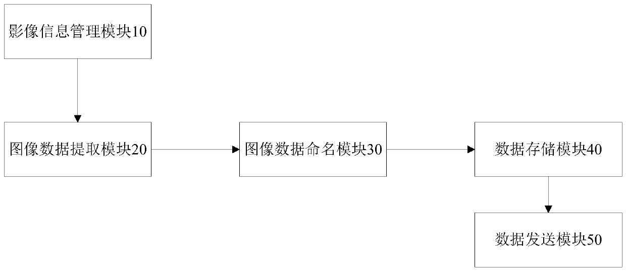
[0029] figure 1 It shows a schematic structural diagram of a system for estimating the phase of a scanning sequence based on DICOM image information according to Embodiment 1 of the present invention; figure 1 As shown, the system includes: image information management module 10, image data extraction module 20, image data naming module 30, data storage module 40 and data sending module 50, wherein,

[0030] The image information management module 10 is connected to the image data extraction module 20, and is used to transmit the patient's DICOM image to the image data extraction module 20 through the DICOM protocol when the patient scans the image examination;

[0031] The image information management module can be a RIS (Radiology Information System) system, and the image inspection can be CT\MR\DR, etc.

[0032] The image data extraction module 20 is connected with the image information management module 10 and the image data naming module 30 respectively, and is used to i...

Embodiment 2

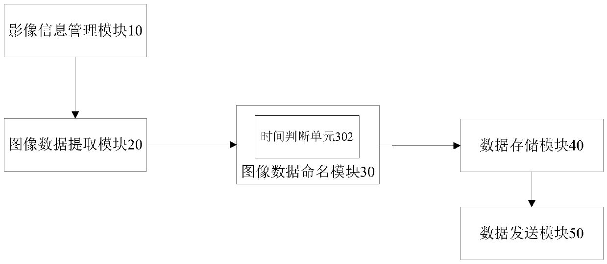
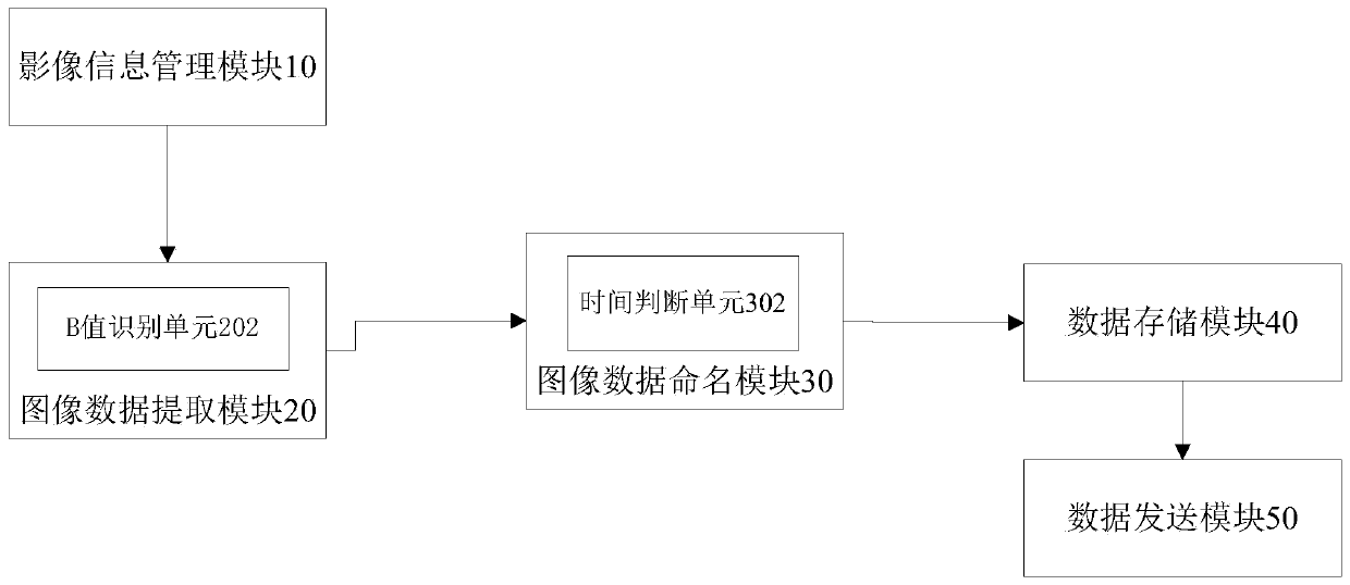
[0045] figure 2 It shows a schematic structural diagram of a system for estimating the phase of a scanning sequence based on DICOM image information according to Embodiment 2 of the present invention; figure 2 As shown, the image data naming module 30 also includes a time judging unit 302, which is used to judge the time length of the scan time data, and judge the phase clinical meaning corresponding to the scan time data based on the time length, examination site data, and spatial position data, Set the name of the DICOM image group according to the clinical meaning of the phase.

[0046] Taking liver CT enhanced scan as an example, after the forearm vein contrast agent is injected, after 15-25 seconds, this time length, combined with the inspection site and spatial position data, is judged as the hepatic artery phase; after 40-50 seconds, this time length , and combined with the inspection site and spatial location data, it is judged as the portal period; after about 10 m...

Embodiment 3

[0049] image 3 It shows a schematic structural diagram of a system for estimating the phase of a scanning sequence based on DICOM image information according to Embodiment 3 of the present invention, as shown in image 3 As shown, when the DICOM image sequence is a DWI sequence, the image data extraction module 20 also includes a B value identification unit 202, which is used for sequence description and B value or spatial position data and scanning time data based on the sequence description in the DICOM image header file information Order and sequence description of DICOM images, determine the B value of DICOM images, and attribute DICOM images with the same B value to the same DICOM image group.

[0050] For example: taking abdominal and pelvic scans as an example, DWI sequence images will have one or more B values, generally 0, 800, 1000, 1200, 1400 and so on. For the judgment of the cancer focus area, the DICOM image group with the highest B value is generally selected,...


PUM


Abstract

The invention provides a system for deducing scanning sequence phase on the basis of DICOM image information, wherein the system includes an image information management module used for transmitting the DICOM image of a patient to an image data extraction module; the image data extraction module can extract a sequence, containing a phase data, in the DICOM image sequence on the basis of inspectionarea data, spatial position data and scanning time data, and then divide the sequence into N DICOM image groups, the images in each of the DICOM image group comprising the same phase data. An image data naming module sets the name of each DICOM image group according to the clinical meanings of the phase on the basis of the sequences of the inspection area data, spatial position data and scanningtime data; a data sending module searches a DICOM image group matched with an AI diagnosis model on the basis of the inspection number of the patient and the name of the DICOM image group and sends the DICOM image group to the AI diagnosis model. The invention further provides a method for deducing scanning sequence phase on the basis of DICOM image information. The system and method are used forimproving diagnosis quality and increasing report writing efficiency.

Description

technical field [0001] The present invention relates to the field of medical information, more specifically, to a system and method for estimating the phase of a scanning sequence based on DICOM image information. Background technique [0002] Contrast imaging is often required during imaging examinations. By observing the filling of target organs in different stages (terms) of contrast imaging, it is helpful to distinguish different types of diseases or judge the severity of diseases. In the prior art, whether it is CT or MR (Nuclear Magnetic), during the scanning process, the machine only records parameters such as the scanned part and time interval, and cannot intelligently judge the phase of each image sequence. This judgment process is In the process of reading the film, the doctor used his own knowledge to judge, but it was not recorded and marked the image sequence. Due to the same scanning scheme and the same part, there may be many image AI (Artificial Intelligence...

Claims


Application Information

Patent Timeline
Patent Type & Authority Applications(China)
IPC IPC(8): G16H30/20G16H50/20
CPCG16H30/20G16H50/20
Inventor 王博李真林刘宇轩邓悟宋伟威
Owner 王博
Why Eureka
Try Eureka