Method for infecting porcine small intestinal mucosa epithelial cell line with porcine epidemic diarrhea virus

A technology of porcine epidemic diarrhea and epithelial cells, applied in the direction of gastrointestinal cells, epidermal cells/skin cells, viruses, etc., can solve problems such as damage to suckling piglets, economic losses in the pig industry, and difficulties in the epithelial cell line of the small intestinal mucosa of pigs

Pending Publication Date: 2020-07-28
ANHUI AGRICULTURAL UNIVERSITY
View PDF3 Cites 2 Cited by
  • Summary
  • Abstract
  • Description
  • Claims
  • Application Information

AI Technical Summary

Problems solved by technology

Pigs of all ages are susceptible, but suckling piglets suffer the most, and the mortality rate can be as high as 100%, which has caused serious economic losses to the pig industry in

Method used


Image

  • Method for infecting porcine small intestinal mucosa epithelial cell line with porcine epidemic diarrhea virus
  • Method for infecting porcine small intestinal mucosa epithelial cell line with porcine epidemic diarrhea virus
  • Method for infecting porcine small intestinal mucosa epithelial cell line with porcine epidemic diarrhea virus

Examples


Embodiment 1

[0039] 1. Method for Infecting Porcine Small Intestinal Epithelial Cell Line with Porcine Epidemic Diarrhea Virus

[0040] 1.1 Use a bottom area of ​​25cm 2 The IEC cell line was cultured in the cell flask, and the volume of the culture medium in the culture was 5 mL. When the cells were covered with a single layer, the culture medium was discarded. Second-rate. Add 1 mL of porcine epidemic diarrhea virus solution containing trypsin 10 μg / mL to each bottle, and place at 37 °C in 5% CO 2 Adsorption in the incubator for 90min. After the adsorption is completed, the liquid is discarded, and DMEM liquid medium containing 5 μg / mL trypsin is added for cultivation and observation. Cytopathy appeared at 36h. It can be seen that the cells fall off and become round, and the intercellular space becomes larger, realizing the in vitro cultivation of porcine epidemic diarrhea virus. The collected cell cultures were repeatedly frozen and thawed three times in a -80°C refrigerator to pre...

Embodiment 2

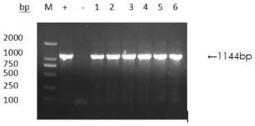
[0063] Real-time fluorescent quantitative detection of the virus copy number of the original virus liquid and the copy number of the 50th generation virus liquid in Example 1, and the RNA was extracted by a conventional extraction method for extracting viral RNA.

[0064] The cDNA after RNA reverse transcription was subjected to PCR reaction to amplify the S gene, (SEQ ID No. 3) upstream primer F: GCAGATTTAGAGCAGCGTTCA, (SEQ ID No. 4) downstream primer R: TAATCAACCAAACCCACCAC.

[0065] The amplification system is as follows: cDNA 2 μL, SYBR GreenMix 10 μL, upstream primer 0.4 μL, downstream primer 0.4 μL, add deionized water to 20 μL, and mix well. The primer concentration was 10 μM.

[0066] The PCR amplification program was as follows: pre-denaturation at 95°C for 15 min; denaturation at 95°C for 10 s, annealing at 61.3°C for 30 s, extension at 72°C for 30 s, and 40 cycles.

[0067] Through the fluorescent quantitative PCR detection method established in the laboratory, the...


PUM

PropertyMeasurementUnit
Copy numberaaaaaaaaaa

Abstract

The invention provides a method for infecting a porcine small intestinal mucosa epithelial cell line with porcine epidemic diarrhea virus, and belongs to the technical field of biotechnology. The method comprises the following steps of mixing small intestinal mucosa epithelial cells with porcine epidemic diarrhea virus liquid, performing adsorbing, removing the liquid, mixing a left substance witha liquid culture medium, and carrying out culturing so as to achieve infection of porcine small intestinal mucosa epithelial cell line by the porcine epidemic diarrhea virus. By adopting the method for culturing the porcine epidemic diarrhea virus in vitro provided by the invention, the porcine epidemic diarrhea virus can be cultured in vitro on the porcine small intestinal mucosa epithelial cellline, and the problem that the porcine epidemic diarrhea virus is difficult to culture on an in vitro target cell is solved; in addition, the copy number of the virus is increased from 106.26 copies/[mu]L to 107.51 copies/[mu]L in the culture process, so that a proper biological material is provided for researching the pathogenesis of the porcine epidemic diarrhea virus.

Description

technical field [0001] The invention belongs to the technical field of biotechnology, and in particular relates to a method for infecting porcine small intestinal mucosal epithelial cell lines with porcine epidemic diarrhea virus. Background technique [0002] Porcine Epidemic Diarrhea Virus (Porcine Epidemic Diarrhea Virus, PEDV) is a member of the Coronaviridae Coronaviridae family, which can cause intestinal infectious diseases of pigs characterized by vomiting, diarrhea and dehydration. Pigs of all ages are susceptible, but suckling piglets suffer the most, with a mortality rate of up to 100%, which has caused serious economic losses to the pig industry in my country and the world. It is difficult for PEDV to proliferate in cell lines cultured in vitro, which makes it difficult for PEDV to infect porcine intestinal mucosal epithelial cell lines. Contents of the invention [0003] The purpose of the present invention is to provide a method for culturing porcine epidemi...

Claims


Application Information

Patent Timeline
IPC IPC(8): C12N5/071C12N7/00
CPCC12N5/0625C12N5/0679C12N7/00C12N2770/20041
Inventor 孙裴秦娟占松鹤赵垚王军何长生王震震李杰李郁
Owner ANHUI AGRICULTURAL UNIVERSITY
Why Eureka
Try Eureka