
Organ three-dimensional image reconstruction method, operation navigation method and operation auxiliary system

A three-dimensional image and auxiliary system technology, applied in the field of surgical assistance systems, can solve the problems that the surgical assistance system cannot present stereoscopic three-dimensional images, cannot present stereoscopic three-dimensional images and surgical navigation, and the reconstructed images are inaccurate. The effect of minor surgical wounds, improving surgical precision, and improving medical services

Active Publication Date: 2020-07-28
SUZE TECH (SHENZHEN) CO LTD
View PDF2 Cites 1 Cited by
  • Summary
  • Abstract
  • Description
  • Claims
  • Application Information

AI Technical Summary

Problems solved by technology

[0005] An object of the present invention is to provide a method for reconstructing a three-dimensional image of an organ, which solves the technical problems that the reconstruction method for a three-dimensional virtual image of an organ in the prior art is relatively complicated and the reconstructed image is not very accurate
[0006] Another object of the present invention is to provide a surgical navigation method, which solves the problem that in the prior art, after reconstruction of the three-dimensional virtual image of organs, only two-dimensional images of three-dimensional modeling can be viewed, and three-dimensional three-dimensional images and operations such as surgical navigation cannot be presented. Technical issues with limited role
[0007] Another object of the present invention is to provide a surgical assistance system to solve the problem that the surgical assistance system in the prior art cannot present a three-dimensional three-dimensional image

Method used


Image

  • Organ three-dimensional image reconstruction method, operation navigation method and operation auxiliary system
  • Organ three-dimensional image reconstruction method, operation navigation method and operation auxiliary system
  • Organ three-dimensional image reconstruction method, operation navigation method and operation auxiliary system

Examples


Embodiment 1

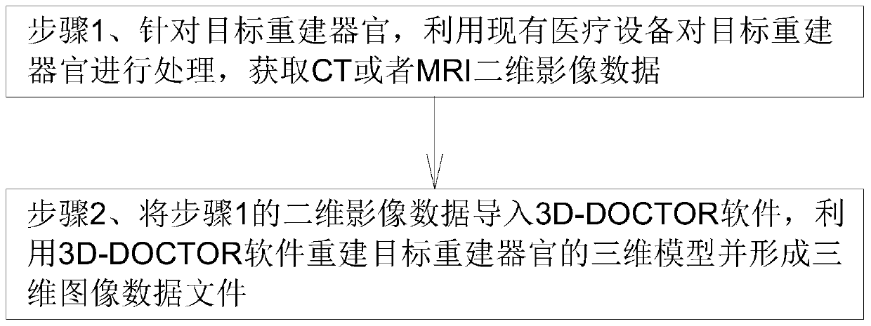
[0044] Such as figure 1 As shown, what this embodiment provides is a method for reconstructing a three-dimensional image of an organ, including the following steps:

[0045] Step 1. For the target reconstructed organ, use existing medical equipment to process the target reconstructed organ, and obtain CT or MRI two-dimensional image data;

[0046] Step 2. Import the two-dimensional image data in step 1 into 3D-DOCTOR software, and use the 3D-DOCTOR software to reconstruct the three-dimensional model of the target reconstructed organ and form a three-dimensional image data file.

[0047]In step 2 of the above steps, after importing the 2D image data in step 1 into the 3D-DOCTOR software, set the name of the target reconstructed organ, draw the area of ​​interest of the target reconstructed organ, set the data format of the 2D image data, and interactively The boundary of the target reconstructed organ is extracted according to the formula, and then the surface is rendered to f...

Embodiment 2

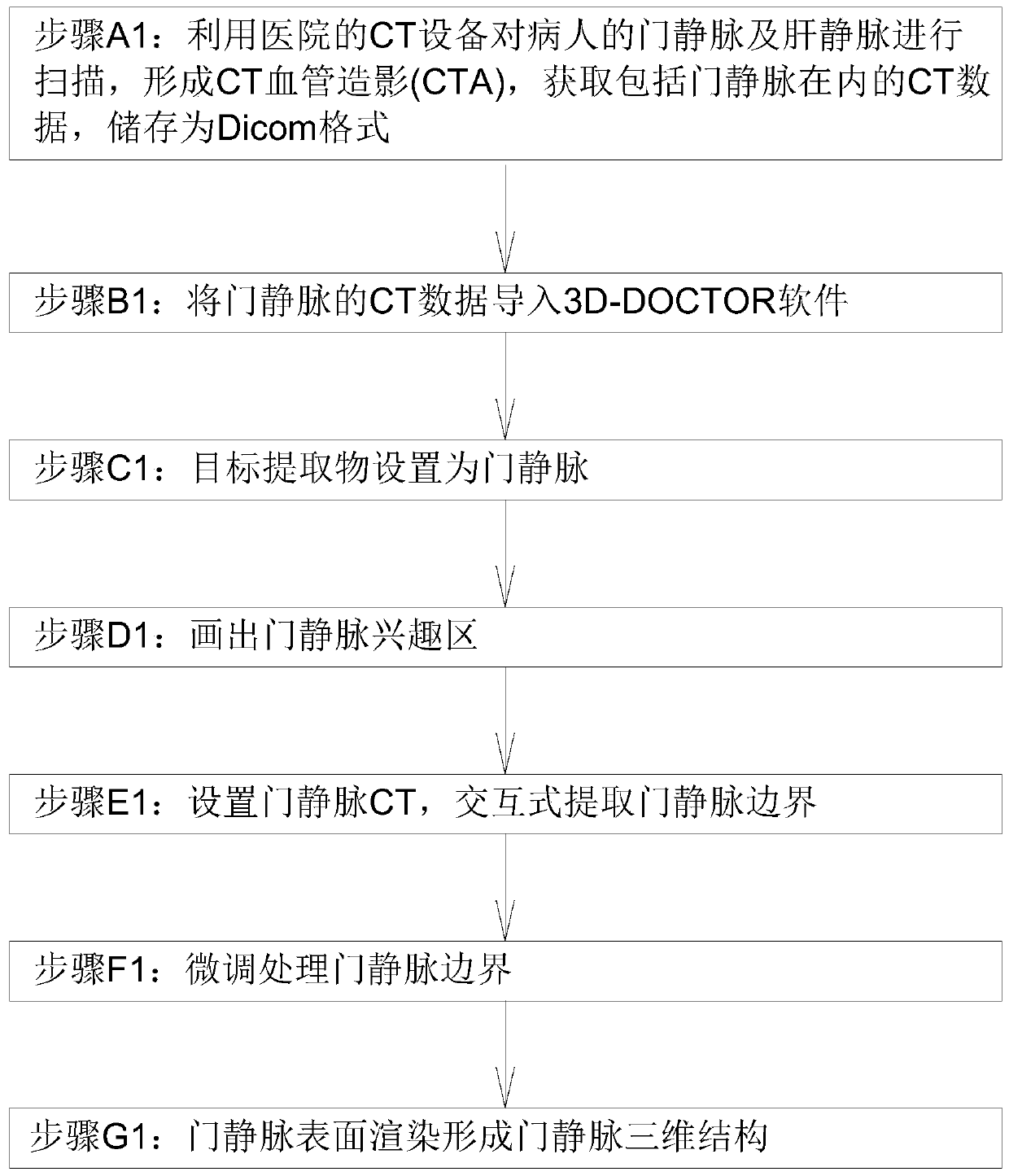
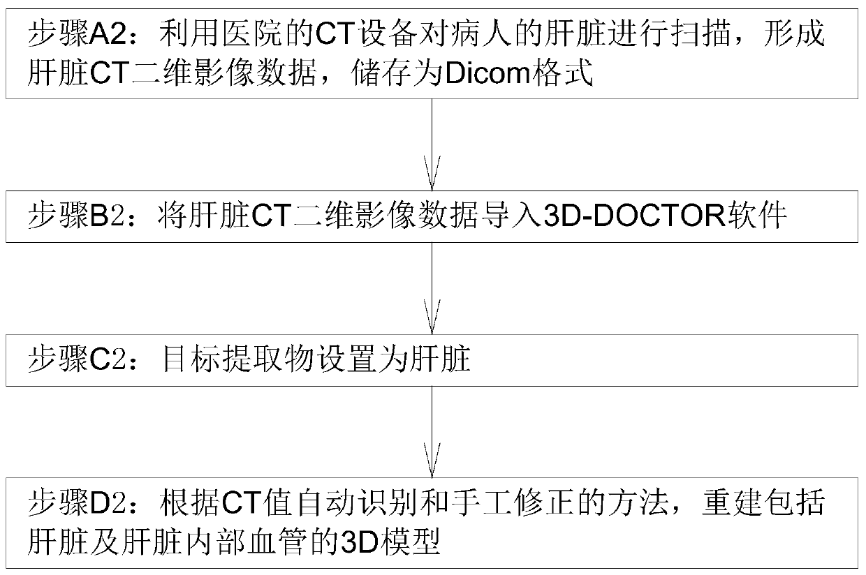
[0057] Such as image 3 , Figure 4 As shown, this embodiment provides a method for reconstructing a three-dimensional image of viscera. The method for reconstructing a three-dimensional image of viscera is similar to the method for reconstructing a three-dimensional image of blood vessels. The liver is used as an example to perform three-dimensional reconstruction of the liver, which specifically includes the following steps:

[0058] Step A2: Use the CT equipment of the hospital to scan the patient's liver to form liver CT two-dimensional image data and store it in Dicom format. The two-dimensional image data is stored in Dicom format with a slice thickness of 2.5mm and an image resolution of 512×512 pixels;

[0059] Step B2: Import liver CT two-dimensional image data into 3D-DOCTOR software, and the software automatically sets the actual scale of the picture;

[0060] Step C2: the target extract is set to liver;

[0061] Step D2: Reconstruct the 3D model including the liv...

Embodiment 3

[0064] Such as Figure 5 to Figure 8 As shown, what this embodiment provides is a method for reconstructing a three-dimensional image of a bone. The method for reconstructing a three-dimensional image of a bone is similar to the method for reconstructing a three-dimensional image of a blood vessel. The following takes the knee joint as an example to perform three-dimensional reconstruction of the knee joint, which specifically includes the following steps:

[0065] Step A3: Use the hospital's nuclear magnetic resonance (MRI) equipment to scan the patient's knee joint to form two-dimensional image data of knee joint MRI. The two-dimensional image data is stored in Dicom format with a slice thickness of 2.5mm and an image resolution of 512×512 pixels ;

[0066] Step B3: Import the knee joint MRI two-dimensional image data into the 3D-DOCTOR software, and the software will automatically set the actual scale of the picture;

[0067] Step C3: The target extract is set to the knee ...


PUM


Abstract

The invention discloses an organ three-dimensional image reconstruction method, an operation navigation method and an operation auxiliary system, and the organ three-dimensional image reconstruction method comprises the following steps: 1, processing a target reconstruction organ through employing existing medical equipment, and obtaining CT or MRI two-dimensional image data; 2, importing the two-dimensional image data in the step 1 into 3D-DOCTOR software, reconstructing a three-dimensional model of a target reconstructed organ by utilizing the 3D-DOCTOR software, and forming a three-dimensional image data file. Existing medical equipment is used for processing a target reconstructed organ. Two-dimensional image data of CT or MRI are acquired, then the two-dimensional image data are imported into 3D-DOCTOR software, the 3D-DOCTOR software is utilized to reconstruct a three-dimensional image of a target reconstructed organ and form a three-dimensional image data file, and the method issimple and accurate in data.

Description

Technical field: [0001] The invention relates to an organ three-dimensional image reconstruction method, an operation navigation method and an operation auxiliary system. Background technique: [0002] Usually, medical imaging equipment only obtains two-dimensional tomographic images of the human body, which is not conducive to people's acquisition of the relative spatial position relationship between various tissues, and cannot meet the requirements for accurate analysis of data. Three-dimensional reconstruction can extract information from image files to create a 3D model, which can display human organs and lesions in three-dimensional form, providing useful visual information for doctors' clinical diagnosis and treatment. Three-dimensional reconstruction technology is the process of recreating three-dimensional images of tissues or organs in the human body based on two-dimensional image slices obtained by CT, MRI, etc., using computer graphics, image processing, and relev...

Claims


Application Information

Patent Timeline
Patent Type & Authority Applications(China)
IPC IPC(8): G06T17/00A61B34/10
CPCG06T17/00A61B34/10A61B2034/105Y02A90/10
Inventor 罗创新黄从云杜晓红杨晓芳
Owner SUZE TECH (SHENZHEN) CO LTD
Learn More