
Eureka
For R&D, Eureka makes reading and utilizing patents & technical documents easy.
Eureka AIR
Designed for self-driven R&D workflows. Generate viable solutions, solve complex R&D challenges, empower your innovation with AI.

Eureka Materials
Designed for material experts only. Revolutionize your material R&D, from search, analyze, to developing new materials.

TechResearch
Generate reliable direction feasibility study reports for your R&D in just a few steps.

TechSeek
Discover and master advanced knowledge NOW. Basics, ideas, possibilities, all at once.

TechMind
As an expert in R&D Theories, TechMind can generates customized viable solutions instantly.

TechRisk
Analyze your overall solution with one click, know your potential R&D risks in advance.

TechMonitor
Get weekly tech updates, stay abreast of the latest tech innovations and key insights.
Ultrasonic endoscope and CT three-dimensional image real-time conversion method and system
A technology of three-dimensional images and two-dimensional images, which is applied in the directions of ultrasonic/sonic/infrasonic diagnosis, acoustic diagnosis, infrasonic diagnosis, etc. It can solve the problem that it is difficult to accurately and clearly collect the location of lesions, and it is impossible to realize the three-dimensional reconstruction of ultrasonic endoscopic images, Unable to obtain pathological diagnosis of biopsy tissue, etc., to achieve the effect of improving the abnormal detection rate, improving the detection accuracy, and reducing the workload
- Summary
- Abstract
- Description
- Claims
- Application Information
AI Technical Summary
Problems solved by technology
Method used
Image

Examples
Embodiment 1
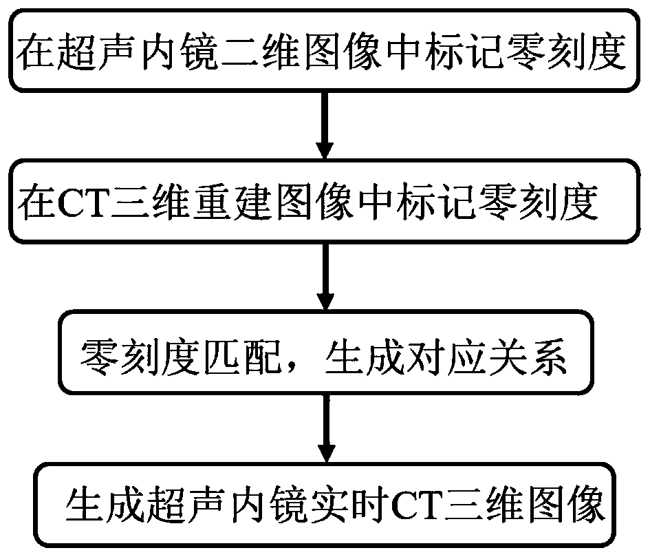
[0025] The present invention provides a method for real-time conversion of ultrasound endoscopy and CT three-dimensional images, including the following steps:
[0026] S1 acquires a two-dimensional image of the ultrasound endoscope, and marks the zero scale in the two-dimensional image.
[0027] Obtain the two-dimensional endoscopic images to be processed, and the doctors with senior professional titles will jointly read the pictures, and screen out the clearer ultrasound endoscopic images from the ascending aorta to the aortic arch. After the screening data, the same two doctors will mark the zero scale , Mark the aorta and the aortic arch as 0 and 1 respectively. The purpose of marking the zero scale of the two-dimensional image is to correspond the zero scale of the two-dimensional image to the zero scale of the three-dimensional image when the two-dimensional image is converted into a three-dimensional image, thereby obtaining the mapping relationship between the two-dimension...
Embodiment 2
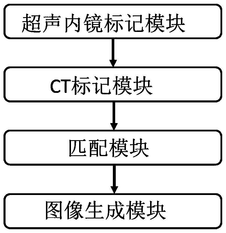
[0041] Based on the same inventive concept, this embodiment discloses a real-time conversion system for ultrasound endoscopy and CT three-dimensional images, including:
[0042] The ultrasound endoscope marking module is used to obtain two-dimensional ultrasound images and mark the zero scale in the two-dimensional images;
[0043] The CT marking module is used to obtain CT 3D reconstruction images and mark the zero scale in the CT 3D reconstruction images;
[0044] The matching module is used to match the zero-scale of the two-dimensional ultrasound image with the zero-scale of the CT three-dimensional reconstructed image, and establish the conversion model of the zero-scale of the two-dimensional ultrasound image and the zero-scale of the CT three-dimensional reconstructed image;
[0045] The conversion module is used to convert the zero scale of the two-dimensional ultrasound image into the CT three-dimensional reconstructed image through the conversion model, and convert the two-di...
PUM
 Abstract
Description
Claims
Application Information
 - R&D Engineer
- R&D Manager
- IP Professional
- Industry Leading Data Capabilities
- Powerful AI technology
- Patent DNA Extraction
Browse by: Latest US Patents, China's latest patents, Technical Efficacy Thesaurus, Application Domain, Technology Topic, Popular Technical Reports.
© 2024 PatSnap. All rights reserved.Legal|Privacy policy|Modern Slavery Act Transparency Statement|Sitemap|About US| Contact US: help@patsnap.com