Cervical cancer lesion diagnosis method fusing multi-modal prior pathological depth characteristics

A technology of deep features and diagnostic methods, applied in the field of medical image processing, can solve problems such as poor diagnostic accuracy of cervical cancer lesions, and achieve the effect of improving efficiency and accuracy

Active Publication Date: 2020-08-04
HUAQIAO UNIVERSITY +1
View PDF3 Cites 10 Cited by
  • Summary
  • Abstract
  • Description
  • Claims
  • Application Information

AI Technical Summary

Problems solved by technology

The current SOTA algorithm generally adopts the fusion method of image data and non-image data (T.Xu, H.Zhang, X.Huang, S.Zhang, and D.Metaxas.Multimodal deep learning for cervical dysplasia diagnosis[C].MICCAI,2016 , 9901:115~123.), there are also related studies that combine acetowhite and iodine-treated images with target detection algorithms for model training (Chen T, Ma X, Ying X, et al.Multi-Modal Fusion Learning for Cervical Dysplasia Diagnosis[ C].In:2019IEEE 16th International Symposium on Biomedical Imaging (ISBI2019).IEEE,2019.1505~1509), the image segmentation method is also applied to cervical images (Zhang, X.Q., S.G.Zhao. Cervical image classification based on imagesegmentation preprocessing and a CapsNet network model[C].In:InternationalJournal of Imaging Systems and Technology,2019,29(1):19~28.); Although these existing algorithms have achieved certain results, due to the deep learning algorithm is not fully utilized The feature correlation of the original image (without drug treatment), vinegar-white processed image, and iodine-processed image makes the accuracy of cervical cancer diagnosis still not good.

Method used


Image

  • Cervical cancer lesion diagnosis method fusing multi-modal prior pathological depth characteristics
  • Cervical cancer lesion diagnosis method fusing multi-modal prior pathological depth characteristics
  • Cervical cancer lesion diagnosis method fusing multi-modal prior pathological depth characteristics

Examples


Embodiment Construction
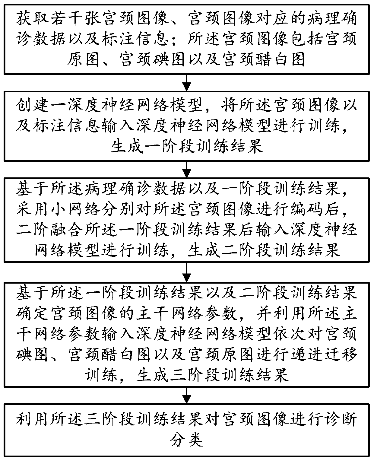
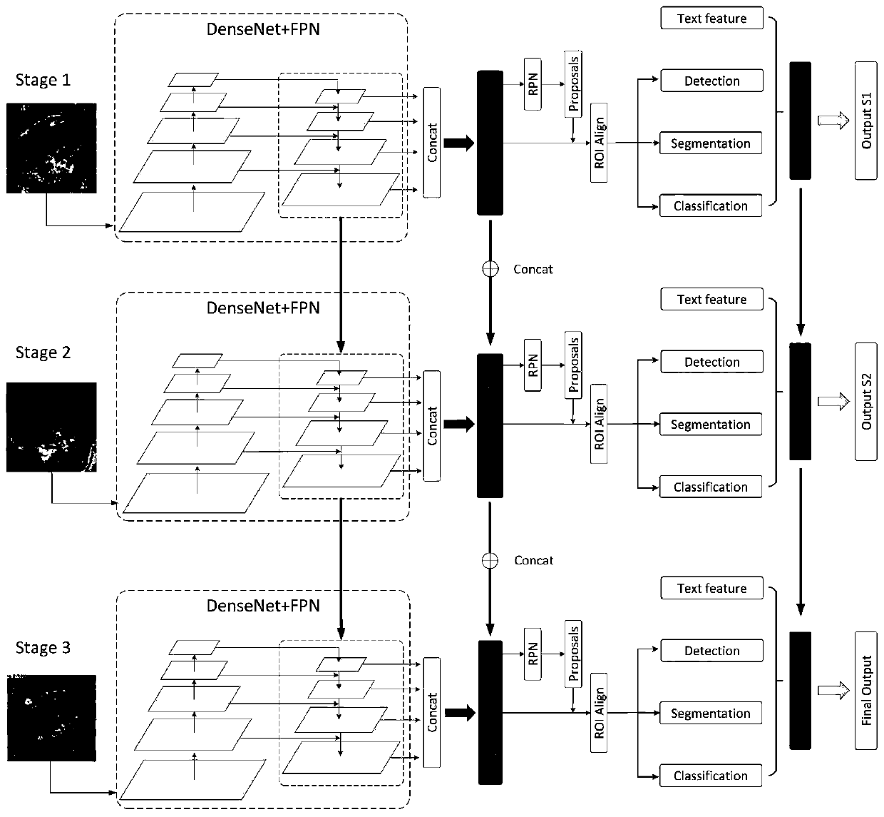
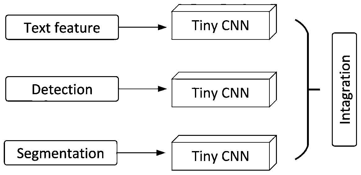
[0043] The general idea of ​​the technical solution in the embodiment of the present application is as follows: combine three kinds of pathological feature images (cervix original image, cervical iodine image and cervical vinegar white image), adopt the main frame of target detection and segmentation, and then use the second-order fusion pathological diagnosis Multi-modal data fusion training was carried out on the data; the three pathological feature images were trained in three stages, and progressive migration training and feature cascade were applied in different stages of training to strengthen the correlation between pathologies, and finally the original image of the cervix (not Accurate diagnosis in drug-treated).

[0044] Please refer to Figure 1 to Figure 4 As shown, a preferred embodiment of a method for diagnosing cervical cancer lesions that combines multimodal prior pathological depth features of the present invention includes the following steps:

[0045] Step ...


PUM


Abstract

The invention provides a cervical cancer lesion diagnosis method fusing multi-modal prior pathological depth features in the field of medical image processing. The method comprises the following steps: step S10, acquiring a cervical image, pathological definite diagnosis data and annotation information; s20, inputting the cervix uteri image and the annotation information into a deep neural networkmodel for training, and generating a first-stage training result; s30, based on the pathology definite diagnosis data and the first-stage training result, coding the cervix uteri image by adopting asmall network, performing second-stage fusion on the first-stage training result, inputting the first-stage training result into a deep neural network model for training, and generating a second-stagetraining result; s40, determining backbone network parameters, inputting the backbone network parameters into the deep neural network model to perform progressive migration training on the cervical image, and generating a three-stage training result; and S50, carrying out diagnosis classification on the cervical images by utilizing a three-stage training result. The method has the advantage thatthe accuracy and efficiency of cervical cancer lesion diagnosis are greatly improved.

Description

technical field [0001] The invention relates to the field of medical image processing, in particular to a method for diagnosing cervical cancer lesions that integrates multimodal prior pathological depth features. Background technique [0002] With the development of science and technology, the advancement of data imaging technology has brought great convenience to medicine, and more and more high-quality images provide a data basis for the analysis and diagnosis of diseases. As one of the early screening methods for cervical cancer, digital colposcopy has been highly recognized all over the world and has become an indispensable technology for the prevention of cervical cancer. Digital colposcopy is a low-cost technology that is becoming more and more mature. Colposcopy mainly uses an optical microscope to magnify the cervix 10 to 40 times under strong light for visual inspection. Doctors with rich experience usually judge whether there is suspicious cancer. to determine th...

Claims


Application Information

Patent Timeline
Patent Type & Authority Applications(China)
IPC IPC(8): G06T5/50G06T7/00G06T7/11G06T7/40G16H30/40G06K9/62G06N3/04
CPCG06T7/0012G06T5/50G06T7/11G06T7/40G16H30/40G06T2207/20016G06N3/045G06F18/214
Inventor 骆炎民林躬耕张涛
Owner HUAQIAO UNIVERSITY
Why Eureka
Try Eureka