Supercharge Your Innovation With Domain-Expert AI Agents!

Image processing method and device

An image processing and elastic image technology, applied in the field of image processing, can solve problems such as difficult differential diagnosis, and achieve the effect of improving accuracy

Pending Publication Date: 2020-08-14
SHENZHEN PEOPLES HOSPITAL
View PDF1 Cites 0 Cited by
  • Summary
  • Abstract
  • Description
  • Claims
  • Application Information

AI Technical Summary

Problems solved by technology

In fact, many nodules with posterior echo attenuation are benign lesions, and the sonographic features of benign and malignant nodules with posterior echo attenuation have common characteristics, making it difficult to differentiate diagnosis

Method used


Image

  • Image processing method and device
  • Image processing method and device
  • Image processing method and device

Examples


Embodiment approach

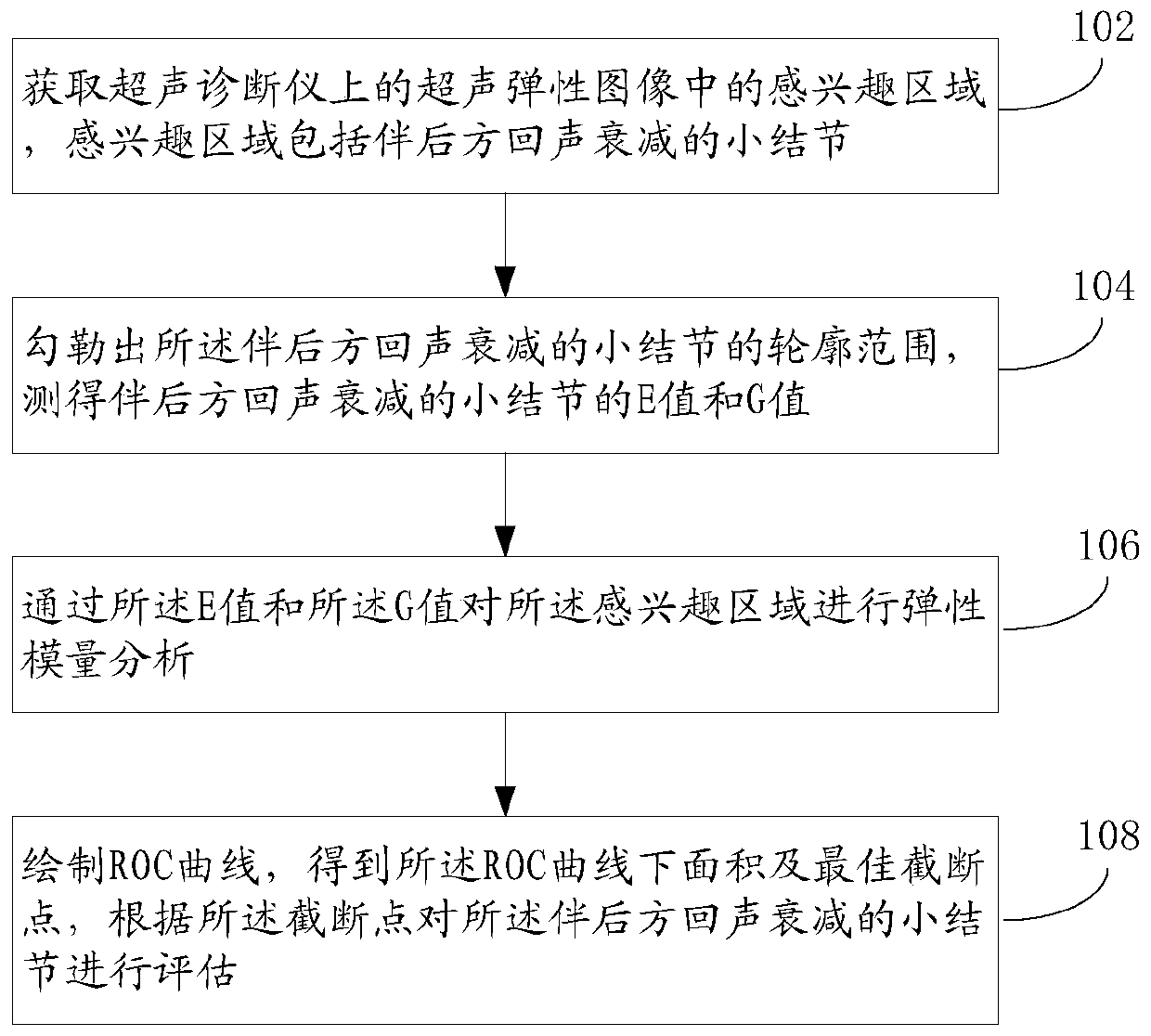
[0053] Such as figure 1 As shown, the image processing method of the present application, an embodiment thereof, comprises the following steps:

[0054] Step 102: Obtain the region of interest in the ultrasound elastic image on the ultrasonic diagnostic instrument, the region of interest includes a small nodule with rear echo attenuation.

[0055] Using Mindray Resona7 ultrasonic diagnostic instrument, 11-3M linear array probe, frequency range 3-11MHz. On the same day, routine ultrasonography was first performed, and the location, size, shape, boundary, echo, calcification, rear echo, and blood flow characteristics of the nodule were observed and recorded. Then turn on the ultra-wide beam tracking technology and enter the elastic STE imaging mode to ensure that all the maximum sections of nodules enter the ROI (region of interest) frame, and turn on the credibility map to help observe the stability of elastic motion and obtain the shearing of nodules. Ultrasound elasticity i...

Embodiment 2

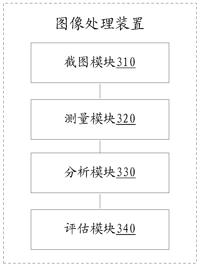
[0073] Such as image 3 As shown, the image processing device of the present application, an implementation manner thereof, may include a screenshot module 310 , a measurement module 320 , an analysis module 330 and an evaluation module 340 .

[0074] The screenshot module 310 is used to obtain the region of interest in the ultrasound elastic image on the ultrasonic diagnostic instrument, and the region of interest includes a small nodule with rear echo attenuation;

[0075] The measurement module 320 is used to outline the outline range of the small nodules with rear echo attenuation, and measure the E value and G value of the small nodules with rear echo attenuation;

[0076] The analysis module 330 is used to analyze the elastic modulus of the region of interest through the E value and the G value;

[0077] The evaluation module 340 is configured to draw the ROC curve, obtain the area under the ROC curve and the optimal cut-off point, and evaluate the small nodules with re...

Embodiment 3

[0096] An image processing device may include a memory and a processor.

[0097] memory for storing programs;

[0098] The processor is configured to implement the method in the first embodiment by executing the program stored in the memory.

[0099]Those skilled in the art can understand that all or part of the functions of the various methods in the foregoing implementation manners can be realized by means of hardware, or by means of computer programs. When all or part of the functions in the above embodiments are implemented by means of a computer program, the program can be stored in a computer-readable storage medium, and the storage medium can include: read-only memory, random access memory, magnetic disk, optical disk, hard disk, etc., through The computer executes the program to realize the above-mentioned functions. For example, the program is stored in the memory of the device, and when the processor executes the program in the memory, all or part of the above-ment...


PUM


Abstract

The invention relates to an image processing method and device. The image processing method comprises the following steps of obtaining regions of interest in ultransonic elastic images on an ultrasonic diagnostic apparatus, wherein the regions of interest comprise a small nodule with rear echo attenuation; outlining the outline range of the small nodule with rear echo attenuation, and measuring anE value and a G value; performing elastic modulus analysis on the regions of interest through the E value and the G value; drawing an ROC curve to obtain ROC area under curve and a best cut off point; and according to the cut off point, performing assessment on the small nodule with rear echo attenuation. According to the image processing method disclosed by the embodiment of the application, inreal-time shear wave elastic imaging, the shear modulus E value and G value are utilized to distinguish the small nodule with rear echo attenuation, the E value and the G value of benign nodules and malignant nodules in the mammary gland with rear echo attenuation are measured, and tissue morphological structure information provided by ultransonic is combined with elastic E imaging hardness information, so that tumor diagnosis accuracy is improved.

Description

technical field [0001] The present application relates to image processing, and in particular to an image processing method and device. Background technique [0002] In the Breast Imaging Reporting and Data System (BIRADS) dictionary, "rear echo attenuation" is one of the words used to describe malignant breast tumors. Classical ultrasound medical books also classify rear echo attenuation as an ultrasound feature of malignant breast lesions. Therefore, in the past Sonographers who see nodules with such characteristics will classify them as breast cancer. In fact, many nodules with posterior echo attenuation are benign lesions, and benign and malignant nodules with posterior echo attenuation have common features on sonograms, making it difficult to differentiate and diagnose. Contents of the invention [0003] The present application provides an image processing method and device. [0004] According to the first aspect of the present application, the present application p...

Claims


Application Information

Patent Timeline
Patent Type & Authority Applications(China)
IPC IPC(8): A61B8/00A61B8/08
CPCA61B8/5215A61B8/485A61B8/0825A61B8/469
Inventor 罗慧林琪肖晓君
Owner SHENZHEN PEOPLES HOSPITAL
Features
  • R&D
  • Intellectual Property
  • Life Sciences
  • Materials
  • Tech Scout
  • Unparalleled Data Quality
  • Higher Quality Content
  • 60% Fewer Hallucinations
Learn More