Nephroscope used for percutaneous nephrolithotomy
A nephroscopic and surgical technology, applied in the field of percutaneous nephroscopy and new medical tools, can solve the problems of increasing the difficulty of surgical operation, limited viewing range of the viewing angle, and impracticality, so as to reduce the residual stone and improve the stone crushing effect. , the effect of improving the crushing efficiency
- Summary
- Abstract
- Description
- Claims
- Application Information
AI Technical Summary
Problems solved by technology
Method used
Image
Examples
Embodiment 1
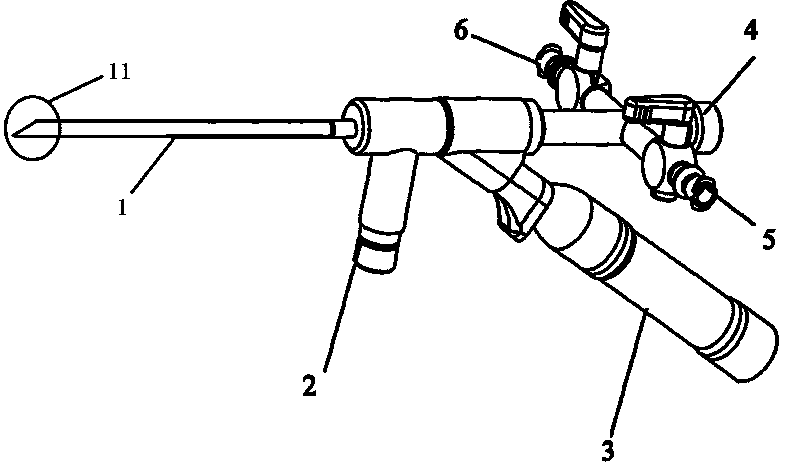
[0026] Such as figure 1 As shown, a nephroscope for percutaneous nephroscopic surgery includes a rigid endoscope 1 , a cold light source input terminal 2 , an image data output terminal 3 , an instrument channel 4 , a water inlet channel 5 and a water outlet channel 6 . The cold light source input end 2 and the image data output end 3 are located on the same side of the central axis of the rigid endoscope 1. The cold light source input end 2 is designed at 90 degrees to the central axis, and the image data output end 3 is designed at a 45-degree angle to the central axis. It has a "gun-like" structure, which effectively enhances the operator's grasp and stability. The rigid endoscope 1 is linear, the front end 11 of the rigid endoscope is a non-curved end, and a liquid channel, a laser device and an imaging device are arranged inside the rigid endoscope 1 .
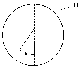
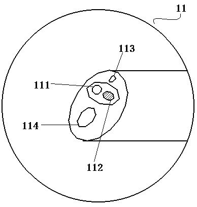
[0027] Such as figure 2 , image 3 As shown, the angle between the plane where the port of the front end 11 of the ...
Embodiment 2
[0031] A nephroscope for percutaneous nephroscopic surgery, its structure is as described in Embodiment 1, the difference is that the port of the front end 11 of the rigid endoscope 1 is located between the plane of the port and the radial plane of the rigid endoscope 1 The angle between them is θ=40°, and the viewing angle of the lens is 40°.
Embodiment 3
[0033] A nephroscope for percutaneous nephroscopic surgery, its structure is as described in Embodiment 1, the difference is that the port of the front end 11 of the rigid endoscope 1 is located between the plane of the port and the radial plane of the rigid endoscope 1 The angle between them is θ=50°, and the viewing angle of the lens is 50°.
PUM
 Abstract
Description
Claims
Application Information
 - R&D
- Intellectual Property
- Life Sciences
- Materials
- Tech Scout
- Unparalleled Data Quality
- Higher Quality Content
- 60% Fewer Hallucinations
Browse by: Latest US Patents, China's latest patents, Technical Efficacy Thesaurus, Application Domain, Technology Topic, Popular Technical Reports.
© 2025 PatSnap. All rights reserved.Legal|Privacy policy|Modern Slavery Act Transparency Statement|Sitemap|About US| Contact US: help@patsnap.com


