Medical image X-ray film developing device

A medical image and film technology, applied in optics, photography, photosensitive materials, etc., can solve problems such as increased work intensity, low efficiency, and shadow liquid splashing, and achieve the effects of ensuring photocopying efficiency, increasing film processing quality, and novel design

Inactive Publication Date: 2020-09-18
刘浩
View PDF1 Cites 0 Cited by
  • Summary
  • Abstract
  • Description
  • Claims
  • Application Information

AI Technical Summary

Problems solved by technology

[0004] The current X-ray film development is to fix the film by clips, the operation is more troublesome, the efficiency is low, and the fixing effect is difficult to guarantee, it is easy to make the film fall off in the clinical radiology department, and it is often necessary to wash negative films of various specifications. X-ray films need to be manually shaken in the film tank to fully wash out the X-ray film, which not only increases the work intensity, but also causes the film to splash on the body, seriously affecting the health of medical staff. good health, practicality
[0005] Poor, which reduces the efficiency of film production, which is not conducive to popularization and use
[0006] Chinese patent (notification number: CN207473295U) discloses a medical image X-ray film developing and drying device, including a support table, a drive shaft is provided in the middle of the support table, a drive gear is installed in the middle of the drive shaft, and the upper end of the support table is The first L-shaped bracket is i

Method used


Image

  • Medical image X-ray film developing device
  • Medical image X-ray film developing device
  • Medical image X-ray film developing device

Examples


Embodiment 1

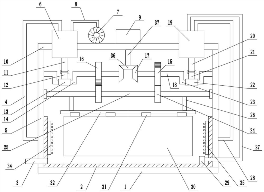
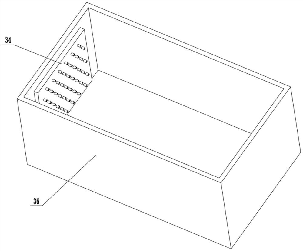
[0030] refer to Figure 1~3, in an embodiment of the present invention, a medical image X-ray film developing device, comprising a fixed base plate 1, a photoprint tank 2 in the middle of the upper end of the fixed base plate 1, a drying chamber 34 is installed on the left side of the inner wall of the photoprint tank 2, and the inner wall of the photoprint tank 2 The liquid spray chamber 35 is installed on the right side, and the bottom of the photocopy tank 2 is also equipped with a liquid discharge pipe 3, which is used to realize the discharge of the liquid after the photocopying is completed. A support frame 5 is installed on both sides of the upper end of the fixed bottom plate 1, and a top plate is installed on the top of the support frame 5. 10. A drying box 6 is installed on the left side of the upper end of the top plate 10. One end of the drying box 6 is connected to a drying chamber 34 through the drying pipe 4, and the other end is connected to the induced draft fa...

Embodiment 2

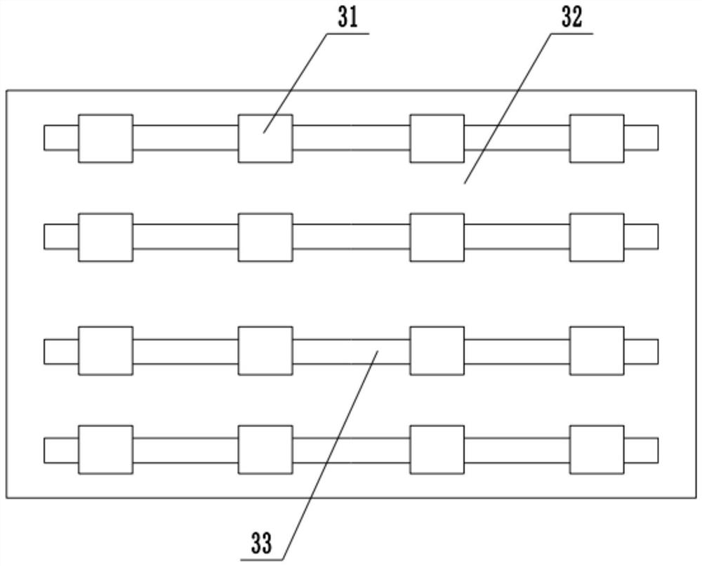
[0032] In another embodiment of the present invention, the difference between this embodiment and the above-mentioned embodiment is that a plurality of sliding grooves 33 are arranged in the middle part of the lower end of the swing plate 32, and a plurality of sliding blocks 31 are installed inside the sliding grooves 33, and the sliding blocks The X-ray film 30 is installed detachably at the lower end of the 31, and some chute 33 and slide block 31 are set to realize the fixation of some X-ray films 30 before and after, which further increases the film processing efficiency.

[0033] In the present invention, when working, the X-ray film 30 is placed at the bottom of the slider 31 for fixing, and at the same time, photoprinting solution is placed inside the photoprinting tank 2, and then the driving motor 9 is started, under the action of the driving gear 36 and the driven gear 17 The rotating shaft 18 is rotated, and then the left gear 16 and the right gear 15 are intermitte...


PUM


Abstract

The invention relates to the technical field of medical equipment. The invention discloses a medical image X-ray film developing device. The device comprises a fixed bottom plate; support frames are mounted on two sides of the upper end of the bottom plate; a top plate is arranged on the top ends of the support frames; a driving motor is mounted in the middle of the top plate; a driving shaft is connected with the lower end of the driving motor; a driving gear is arranged below the driving shaft; driven gears are engaged with two sides of the lower end of the driving gear; rotating shafts aremounted in the driven gear; a left gear is mounted in the right middle of the left rotating shaft; a first crank is mounted in the middle of the left end; a first sliding sleeve is installed in the middle of the first crank; a first connecting rod is installed at the upper end of the first sliding sleeve; a first piston rod is hinged to the upper end of the first connecting rod; the top end of thefirst piston rod is connected with a piston plate arranged in a drying box; a right gear is installed in the middle of the left end of the right rotating shaft. According to the invention, an X-ray film swings back and forth in a photographic printing groove, the film developing quality is improved, and meanwhile the photographic printing efficiency and the drying efficiency after photographic printing are further ensured.

Description

technical field [0001] The invention relates to the technical field of medical equipment, in particular to a medical image X-ray film developing device. Background technique [0002] X-rays are particle streams produced by the transition of electrons in atoms between two energy levels with greatly different energies, and are electromagnetic waves with wavelengths between ultraviolet rays and gamma rays. Its wavelength is very short, between 0.01 and 100 Angstroms. It was discovered by German physicist W.K. Roentgen in 1895, so it is also called Roentgen ray. Rays have high penetrating power and can pass through many substances that are opaque to visible light, such as ink paper, wood, etc. This invisible ray can cause visible fluorescence in many solid materials, sensitize photographic negatives, and ionize air. X-rays were originally used in medical imaging diagnostics and X-ray crystallography. X-rays are also harmful rays such as ionizing radiation. [0003] With the...

Claims


Application Information

Patent Timeline
IPC IPC(8): G03D3/00G03D13/08G03D15/02
CPCG03D3/00G03D13/08G03D15/02
Inventor 刘浩
Owner 刘浩
Why Eureka
Try Eureka