Three-dimensional image detection system and device, imaging method and lung detection device

A three-dimensional imaging and detection system technology, applied in the field of medical imaging, can solve the problems of inconvenient operation, low usage rate, and difficulty in disinfection, and achieve the effects of simple operation, improved utilization rate, and reduced radiation range.

Pending Publication Date: 2020-10-16
SHANGHAI INNOMOTION
View PDF0 Cites 3 Cited by
  • Summary
  • Abstract
  • Description
  • Claims
  • Application Information

AI Technical Summary

Problems solved by technology

[0006] The embodiments of the present application provide a three-dimensional image detection system, device, imaging method, and lung detection device to solve the problem of inconvenient operation, large radiation, low utilization r

Method used


Image

  • Three-dimensional image detection system and device, imaging method and lung detection device
  • Three-dimensional image detection system and device, imaging method and lung detection device
  • Three-dimensional image detection system and device, imaging method and lung detection device

Examples


Embodiment 1

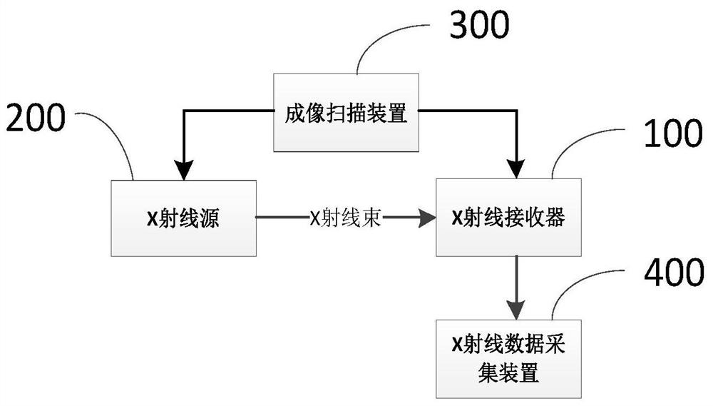
[0052] Reference attachment Figure 1-3 This embodiment provides a three-dimensional image detection system, which includes an X-ray receiver 100 and an X-ray source 200. Wherein, the X-ray source 200 is used to emit X-ray beams, the X-ray receiver 100 is used to receive X-ray beams, and the X-ray receiver 100 in this embodiment is used to receive X-ray beams that penetrate the object to be inspected.

[0053] Further, the three-dimensional image detection system further includes: an imaging scanning device 300 and an X-ray data acquisition device 400.

[0054] The imaging scanning device 300 is configured to transmit the X-ray beam emitted by the X-ray source 200, penetrate the object to be inspected, and aim at the X-ray receiver 100; and then drive the X-ray source 200 to move in the first direction of the object to be inspected while moving around the X-ray The center of the ray source 200 rotates in the first direction to perform angular compensation of the X-ray beam, so that...

Embodiment 2

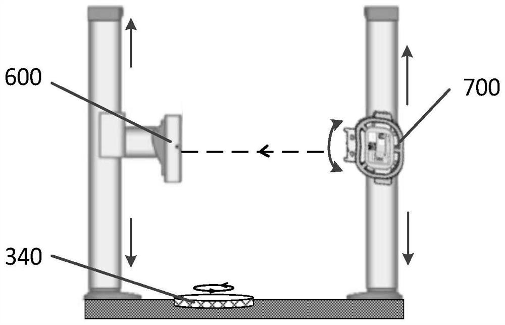
[0072] Reference attachment Figure 4-5 This embodiment provides a three-dimensional image detection device, which adopts the three-dimensional image detection system in the first embodiment, and includes: a supporting assembly 500, a receiving end 600, and a transmitting end 700.

[0073] The receiving end 600 includes an X-ray source 200 for emitting X-ray beams at different angles in the first direction to penetrate the object to be inspected.

[0074] The transmitting end 700 includes an X-ray receiver 100 for receiving X-ray beams transmitted through the object to be detected.

[0075] The receiving terminal 600 and the transmitting terminal 700 are arranged on the supporting device oppositely; among them, the space between the transmitting terminal 700 and the receiving terminal 600 is used as the accommodating area of ​​the detected object, so that the X-ray receiver 100 can receive X-rays penetrating the detected object bundle.

[0076] Attached Figure 4 The three-dimensional...

Embodiment 3

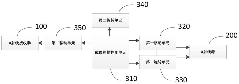
[0087] This embodiment provides an imaging method of the three-dimensional image detection device of the second embodiment. The three-dimensional image detection system includes an imaging scanning device 300 and an X-ray data acquisition device 400. The imaging scanning device 300 includes a first moving unit 320 and a second moving unit 320. The moving unit 350, the imaging scan control unit 310, the second rotating unit 340, and the second moving unit 350.

[0088] The imaging method includes the following steps:

[0089] Step S1: The X-ray beam emitted by the X-ray source 200 is driven to penetrate the object to be inspected and align with the X-ray receiver 100. Step S1 further includes driving the X-ray receiver 100 to move in the first direction of the detected object, and position the detected object to receive the X-ray beam penetrating the detected object.

[0090] Step S2: Drive the X-ray source 200 to move in the first direction of the detected object, and at the same ti...


PUM


Abstract

The invention discloses a three-dimensional image detection system and device, an imaging method and a lung detection device. The three-dimensional image detection system comprises an X-ray receiver,an X-ray source, an imaging scanning device and an X-ray data collection device, wherein an X-ray beam emitted by the X-ray source penetrates through a detected object, and is directed at the X-ray receiver; the X-ray source is driven to move and rotate, so that the X-ray beam penetrates through the detected object at different angles of a first direction; the X-ray receiver and the X-ray source are driven to rotate synchronously in a second direction perpendicular to the first direction, so that the X-ray beam rotates in the second direction perpendicular to the first direction; and the X-raydata collection device is configured to drive the system in a computerized tomography mode, and multiple X-ray projected images are generated in multiple selective positions of the detected object bythe X-ray beam emitted by the X-ray source so that a three-dimensional reconstructed image of the detected object can be generated through processing. By means of a lung imaging system, effects of simple operation, easy carrying, easy protection, low radiation and the like are achieved.

Description

Technical field [0001] The invention relates to medical imaging technology, in particular to a three-dimensional image detection system, device, imaging method and lung detection device. Background technique [0002] With the development of imaging technology, in medicine, more and more medical images are used to discover and diagnose diseases. However, this diagnosis method may cause doctors to misjudge and misdiagnose due to the contrast and resolution of the image itself. At the same time, it is quite difficult and inaccurate to conceive the three-dimensional structure of human organs from a set of two-dimensional images, and may even cause misdiagnosis and cause unimaginable consequences. [0003] In order to improve the scientificity and accuracy of medical diagnosis and treatment planning, it is necessary to process two-dimensional tomographic images, highlight and observe important parts, and shield or delete unnecessary parts. Existing two-dimensional images are converted ...

Claims


Application Information

Patent Timeline
IPC IPC(8): A61B6/00
CPCA61B6/40A61B6/42A61B6/4411A61B6/4435A61B6/4476A61B6/5211
Inventor 王少白皇甫良周武建侯尧兰天吴琳琳
Owner SHANGHAI INNOMOTION
Why Eureka
Try Eureka