Breast tissue elasticity imaging method, device and equipment and medium

An elastography and breast tissue technology, applied in the field of medical image processing, can solve problems such as affecting doctors' clinical diagnosis, small imaging field of view, and inability to accurately locate

Pending Publication Date: 2020-10-23
SHENZHEN UNIV
View PDF13 Cites 0 Cited by
  • Summary
  • Abstract
  • Description
  • Claims
  • Application Information

AI Technical Summary

Problems solved by technology

[0004] However, since the 3D ultrasound images and 2D elastic images need to be acquired by different instruments, the method of viewing the 3D ultrasound grayscale images and 2D elastic images at the same time with two monitors is cumbersome and takes a long time; moreover, the actual The tumor is three-dimensional, and the two-dimensional elastic image cannot truly and comprehensively reflect the state of the entire tumor. If some important image information is not obtained during the examination due to the limitation of the imaging principle, it may affect the doctor's clinical diagnosis. The "fault" corresponding to the 3D ultrasound image corresponding to the 2D elastic image cannot be accurately located only with the naked eye
The method of directly performing three-dimensional elastography with a handheld area array probe requires manual operation for imaging the entire tissue, and the relationship between the obtained fields of view is difficult to standardize, and the field of view is small, much smaller than the imaging field of view of the overall ultrasound image of the tissue.

Method used


Image

  • Breast tissue elasticity imaging method, device and equipment and medium
  • Breast tissue elasticity imaging method, device and equipment and medium
  • Breast tissue elasticity imaging method, device and equipment and medium

Examples


Embodiment 1

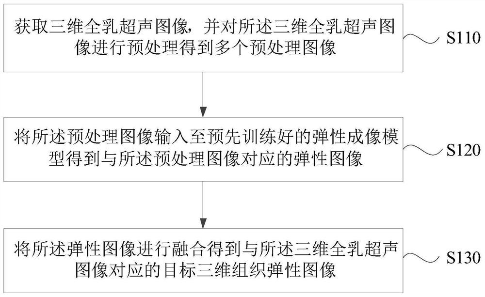
[0048] figure 2 It is a flow chart of the breast tissue elastography method provided in Embodiment 1 of the present invention. This embodiment is applicable to the case of obtaining a three-dimensional breast tissue elastic image. This method can be realized by a breast tissue elastography device configured in a computer device , specifically, it may be implemented through software and / or hardware in the device.

[0049] like figure 2 As shown, the breast tissue elastography method specifically includes:

[0050] S110. Acquire a three-dimensional whole-breast ultrasound image, and perform preprocessing on the three-dimensional whole-breast ultrasound image to obtain a plurality of preprocessed images.

[0051]Wherein, the imaging object of the imaging method is breast tissue. The three-dimensional full-breast ultrasound image is an ultrasound grayscale image obtained by injecting ultrasound beams into the target tissue, obtaining reflected signals and processing the refle...

Embodiment 2

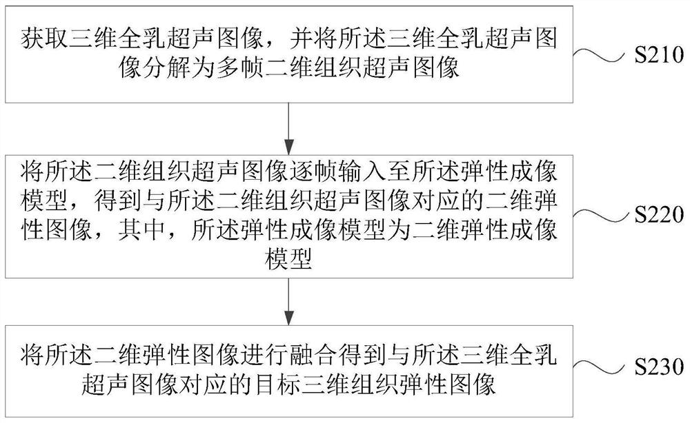
[0060] image 3 The flow chart of the breast tissue elastography method provided by Embodiment 2 of the present invention, on the basis of the above embodiments, this embodiment further illustrates the input of the elastography model as the two-dimensional tissue ultrasound image and the output as the corresponding two-dimensional elastic The condition of the image.

[0061] like image 3 As shown, the breast tissue elastography method specifically includes:

[0062] S210. Acquire a three-dimensional whole-breast ultrasound image, and decompose the three-dimensional whole-breast ultrasound image into multiple frames of two-dimensional tissue ultrasound images.

[0063] In three-dimensional tissue ultrasound imaging, the three-dimensional whole-breast ultrasound image can be translated by a two-dimensional ultrasonic probe at the end of a mechanical arm probe, driven by the mechanical arm. The probe acquires a two-dimensional ultrasound image, and finally the two-dimensional...

Embodiment 3

[0070] Figure 4 The flow chart of the breast tissue elastography method provided by Embodiment 3 of the present invention, on the basis of the above embodiments, this embodiment further illustrates the input of the elastography model as the three-dimensional whole-breast ultrasound image block and the output as the corresponding three-dimensional elastic The case of image blocks.

[0071] like Figure 4 As shown, the breast tissue elastography method specifically includes:

[0072] S310. Acquire a three-dimensional whole-breast ultrasound image, and divide the three-dimensional whole-breast ultrasound image into multiple three-dimensional whole-breast ultrasound image blocks of a preset size.

[0073] In this embodiment, the input of the elastography model is a small block of a three-dimensional whole-breast ultrasound image, therefore, the three-dimensional whole-breast ultrasound image needs to be divided into multiple three-dimensional whole-breast ultrasound image block...


PUM


Abstract

The embodiment of the invention discloses a breast tissue elasticity imaging method, device and equipment and a medium. The method comprises the following steps: acquiring three-dimensional whole milkultrasonic images, and carrying out the preprocessing of the three-dimensional whole milk ultrasonic images to obtain a plurality of preprocessed images; inputting the preprocessed image into a pre-trained elastic imaging model to obtain elastic images corresponding to the preprocessed images; and fusing the elastic images to obtain a target three-dimensional tissue elastic image corresponding tothe three-dimensional whole-emulsion ultrasonic image. According to the technical scheme of the embodiment of the invention, the problems that a three-dimensional whole-emulsion ultrasonic image doesnot contain elastic information and the visual field range is small by directly adopting an area array probe for three-dimensional elastic imaging are solved; and the three-dimensional ultrasonic elastic image can be directly generated according to the three-dimensional ultrasonic grayscale image, and three-dimensional imaging can be performed without secondary image acquisition or manual operation, so that the acquisition efficiency of the three-dimensional elastic image is improved, and disease diagnosis is facilitated.

Description

technical field [0001] Embodiments of the present invention relate to the technical field of medical image processing, and in particular to a breast tissue elasticity imaging method, device, equipment and medium. Background technique [0002] Three-dimensional ultrasound imaging is a technology that combines ultrasound probes with computer processing to obtain three-dimensional image information, in which a series of two-dimensional imaging is performed using a two-dimensional ultrasound probe, and then the two-dimensional images are reconstructed into three-dimensional images. It has the characteristics of comprehensive field of view, accurate positioning, intuitive images, and high degree of image standardization, so it has been widely used, and three-dimensional whole breast ultrasound is one of them. Ultrasonic elastography is a method of inducing tissue deformation by pressurization, shear waves, etc., and imaging the degree of tissue deformation before and after pressu...

Claims


Application Information

Patent Timeline
Patent Type & Authority Applications(China)
IPC IPC(8): A61B8/08A61B8/00
CPCA61B8/0825A61B8/483A61B8/485A61B8/5215A61B8/5223A61B8/5246A61B8/5253
Inventor 高毅王菁宋宏萍
Owner SHENZHEN UNIV
Why Eureka
Try Eureka