Method for measuring fetal corpus callosum volume by using magnetic resonance imaging, and magnetic resonance imaging apparatus

A technology of magnetic resonance imaging and measurement method, which is applied in measurement devices, measurement of magnetic variables, neural learning methods, etc., can solve the problem of inability to accurately calculate the volume of the corpus callosum, inability to provide imaging methods, and inability to provide effective judgment by sonographers basis, etc.

Pending Publication Date: 2020-10-30
SIEMENS HEALTHINEERS LTD +1
View PDF0 Cites 4 Cited by
  • Summary
  • Abstract
  • Description
  • Claims
  • Application Information

AI Technical Summary

Problems solved by technology

[0003] At present, ultrasound images are usually used for fetal screening, but the use of ultrasound images to screen for abnormalities of the corpus callosum requires high requirements for the operator's experience and skills, and ultrasound images cannot accurately calculate the volume of the corpus callosum. It also cannot provide an effective basis for the judgment of the sonographer
Therefore, at present, the abnormality of the fetal corpus callosum is not easy to judge by ultrasound images
[0004] In addition...

Method used


Image

  • Method for measuring fetal corpus callosum volume by using magnetic resonance imaging, and magnetic resonance imaging apparatus
  • Method for measuring fetal corpus callosum volume by using magnetic resonance imaging, and magnetic resonance imaging apparatus
  • Method for measuring fetal corpus callosum volume by using magnetic resonance imaging, and magnetic resonance imaging apparatus

Examples


Embodiment Construction

[0024] In order to make the purpose, technical solution and advantages of the present invention clearer, the following examples are given to further describe the present invention in detail.

[0025] An embodiment of the present invention provides a method for measuring the volume of the fetal corpus callosum by using magnetic resonance imaging. According to the method, the volume of the fetal corpus callosum can be obtained, thereby providing an auxiliary judgment for judging the abnormality of the corpus callosum.

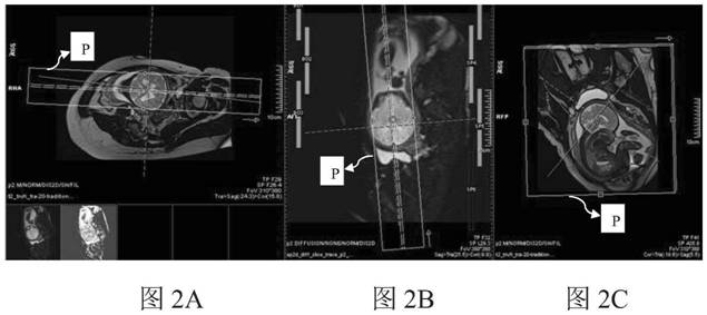
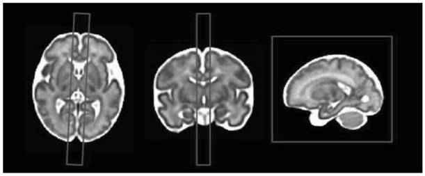
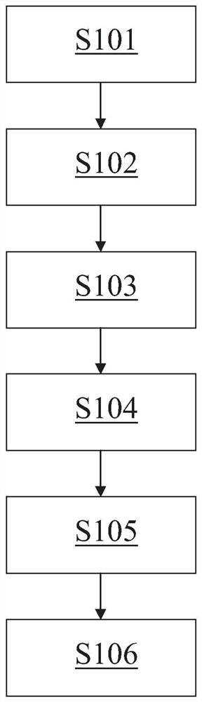
[0026] figure 1 The measurement method of fetal corpus callosum volume using magnetic resonance imaging according to the present invention is shown. Figure 2A The coronal positioning image of the fetus in magnetic resonance imaging and the position of the detection region P (rectangular wire frame in the figure) in the positioning image are shown. Figure 2B A transverse positional scout of the fetus and the position of the detection region P in the scout are s...


PUM


Abstract

The invention provides a method for measuring a fetal corpus callosum volume by using magnetic resonance imaging, and a magnetic resonance imaging apparatus. The method comprises the following steps:acquiring a positioning image of a to-be-detected fetus through magnetic resonance imaging; determining a detection area P according to the position of fetal corpus callosum in the positioning image,wherein the detection area P comprises the fetal corpus callosum; carrying out magnetic resonance scanning on the detection area to obtain a diffusion weighted image of the detection area, wherein a gradient direction during the magnetic resonance scanning only needs to be applied to a direction parallel to the extension direction of the fiber bundles of the corpus callosum; intercepting a fetal head image in the diffusion weighted image; applying a preset threshold to the fetal head image to obtain high signals with brightness greater than a threshold in the image; and carrying out bunching processing on the high signals, taking the maximum bunch in the high signals as corpus callosum, calculating the sum of the sizes of voxels related to the high signal of the maximum bunch, and taking the sum as the volume of the corpus callosum.

Description

technical field [0001] The invention relates to a method for measuring the volume of the fetal corpus callosum by using magnetic resonance imaging, in particular to a fully automatic method for measuring the volume of the fetal corpus callosum and a magnetic resonance imaging device. Background technique [0002] The corpus callosum is a fiber bundle plate composed of fibers that connect the left and right cerebral hemispheres. Dysplasia of the corpus callosum may cause mild mental retardation or mild visual impairment or cross-tactile positioning disorder. Therefore, abnormalities of the corpus callosum should be screened and appropriate measures should be taken during the fetal period Can reduce the probability of abnormalities in newborns. [0003] At present, ultrasound images are usually used for fetal screening, but the use of ultrasound images to screen for abnormalities of the corpus callosum requires high requirements for the operator's experience and skills, and ul...

Claims


Application Information

Patent Timeline
IPC IPC(8): A61B5/055A61B5/00
CPCA61B5/055A61B5/4362A61B5/7267A61B5/0042A61B5/4064A61B2576/026G01R33/5616G01R33/56341A61B2503/02G06N3/08G06V10/28G06V10/25G06V10/50G06V2201/03G06N20/00
Inventor 王光彬钱天翼陈欣孙聪
Owner SIEMENS HEALTHINEERS LTD
Why Eureka
Try Eureka