
A Method for Detection and Classification of Lesion Areas in Clinical Images

A lesion area and classification method technology, applied in the field of clinical image processing, can solve the problems of only considering single modality and not being able to make full use of multimodal information of clinical images, so as to avoid biopsy, avoid manual heuristic learning, and reduce concurrency disease effect

Active Publication Date: 2022-04-01
NANJING TUGE HEALTHCARE CO LTD
View PDF4 Cites 0 Cited by
  • Summary
  • Abstract
  • Description
  • Claims
  • Application Information

AI Technical Summary

Problems solved by technology

However, most current deep learning architectures usually only consider single modality, and cannot make full use of the multimodal information of clinical images.

Method used


Image

  • A Method for Detection and Classification of Lesion Areas in Clinical Images
  • A Method for Detection and Classification of Lesion Areas in Clinical Images
  • A Method for Detection and Classification of Lesion Areas in Clinical Images

Examples


Embodiment Construction

[0059] Embodiments of the present invention will be further described in detail below in conjunction with the accompanying drawings.

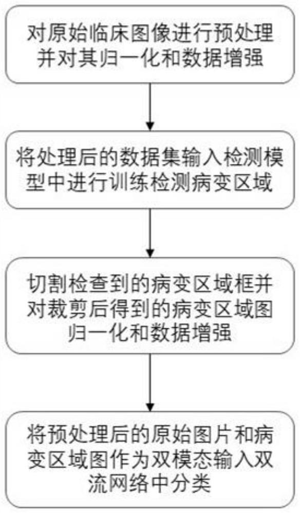
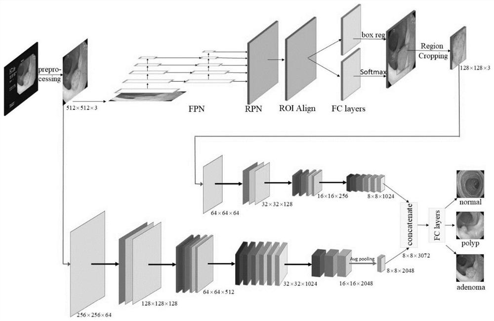
[0060] Such as figure 1 Shown is the flow chart of the clinical image detection and classification method based on multimodal deep learning of the present invention, and the method comprises the following steps:

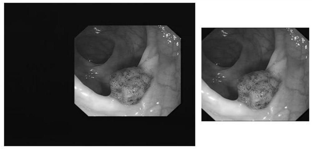
[0061] Step 1. Preprocess the original colonoscopy clinical image, remove redundant information and noise of the colonoscopy clinical image, and normalize and enhance the data to obtain the global image data set;

[0062] Clinical images are obtained by medical imaging systems, and are divided into different types of images according to patient pathology, such as benign and malignant.

[0063] Step 2, inputting the global image data set into a convolutional neural network model (RCNN) based on the detection of target areas in the image for training, and detecting possible lesion areas in the image;

[0064] Step 3. Outline the possi...


PUM


Abstract

The invention discloses a method for detecting and classifying lesion areas in clinical images, which includes preprocessing original clinical images to obtain a global image data set; inputting the obtained global image data set into a convolutional neural network based on detecting target areas in the image The network model is trained to detect the possible lesion area in the image; the detected possible lesion area is framed with a rectangular frame, the rectangular frame area is cut to obtain the lesion area, and the lesion area obtained after cutting is normalized and data enhanced. A local image data set containing the lesion area is obtained; the global image data set and the local image data set containing the lesion area are input into the dual-stream convolutional neural network as bimodal inputs for classification. The method of the present invention better utilizes the multi-modal information of clinical images and combines the global information of the original image with the local information of the lesion area image, thereby improving the accuracy of classification.

Description

technical field [0001] The invention belongs to the technical field of clinical image processing, and in particular relates to a method for detecting and classifying lesion areas in clinical images. Background technique [0002] Tumor is a major killer that endangers life and health, and has a high mortality rate. The clinical manifestations of malignant tumors vary depending on the organ, location, and degree of development. Most malignant tumors have no obvious symptoms in the early stage, and even if they have symptoms, they often have no symptoms. Specific, when specific symptoms appear in patients, the tumor is often already at an advanced stage. A comprehensive analysis of the patient's clinical manifestations and signs is usually performed, combined with laboratory tests, imaging, and cytopathological examinations to make a definite diagnosis, so as to formulate treatment plans and evaluate prognosis. However, there is still a lack of ideal and specific early diagnos...

Claims


Application Information

Patent Timeline
Patent Type & Authority Patents(China)
IPC IPC(8): G06T7/00G06T7/11G06T7/136G06T5/00G06V10/82G06V10/764G06K9/62G06N3/04G06N3/08
CPCG06T7/0012G06T7/136G06T7/11G06N3/08G06T2207/10068G06T2207/20081G06T2207/20084G06T2207/20016G06T2207/30096G06N3/045G06F18/24G06T5/70G06N3/04G06V10/82G06V2201/032G06F18/214
Inventor 汪彦刚王凯妮陈阳周光泉
Owner NANJING TUGE HEALTHCARE CO LTD
Learn More