
Exosome markers for diagnosing lymph node metastasis of invasive breast cancer and application of exosome markers

A technology for lymph node metastasis and infiltration, applied in the field of detection and diagnosis, to achieve high sensitivity, good specificity, and avoid excessive tissue biopsy

Pending Publication Date: 2022-05-03
SHANGHAI FIRST PEOPLES HOSPITAL
View PDF0 Cites 0 Cited by
  • Summary
  • Abstract
  • Description
  • Claims
  • Application Information

AI Technical Summary

Problems solved by technology

[0005] In order to solve the problem in the prior art that there is no high specificity and sensitivity of patient blood exosome molecules for the clinical diagnosis of lymph node metastasis of invasive breast cancer, the present invention provides a set of exosome molecules for the diagnosis of lymph node metastasis of invasive breast cancer Exosome markers and applications, the exosome marker combination has high accuracy in distinguishing lymph node metastasis from patients with invasive breast cancer and no lymph node metastasis, and can be used in the preparation of diagnostic reagents for lymph node metastasis of invasive breast cancer middle

Method used


Image

  • Exosome markers for diagnosing lymph node metastasis of invasive breast cancer and application of exosome markers
  • Exosome markers for diagnosing lymph node metastasis of invasive breast cancer and application of exosome markers
  • Exosome markers for diagnosing lymph node metastasis of invasive breast cancer and application of exosome markers

Examples


Embodiment 1

[0055] The process of screening exosome markers for diagnosing lymph node metastasis of invasive breast cancer will be described in detail below.

[0056] (1) Extraction and identification of exosomes

[0057] 1. Collection of serum samples

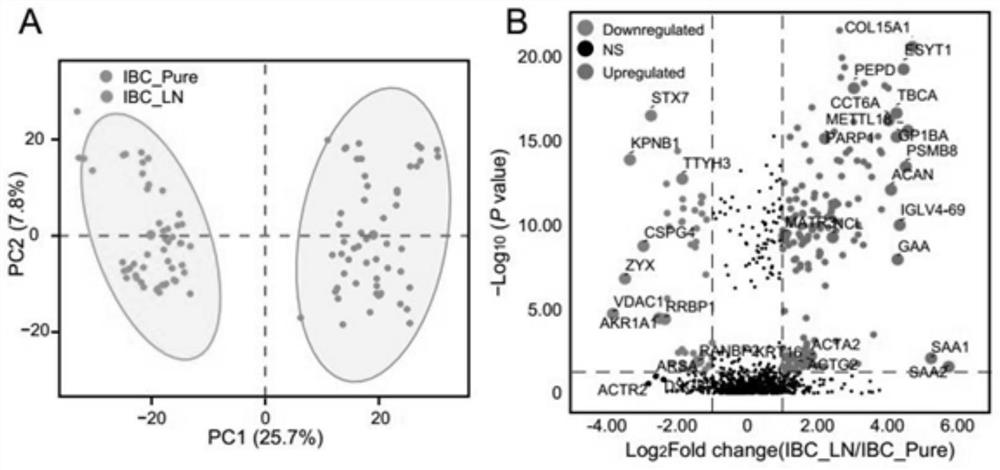
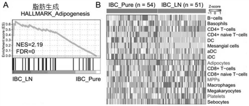
[0058] The serum samples of invasive breast cancer with lymph node metastasis (ibc_ln group) were from 51 patients diagnosed by Shanghai First People's Hospital Affiliated to Medical College of Shanghai Jiao Tong University; Serum samples of in situ invasive breast cancer (ibc_pure group) were also obtained from 54 patients in Shanghai First People's Hospital Affiliated to Medical College of Shanghai Jiao Tong University. These serum samples were placed in the refrigerator at - 80 ℃ for detection.

[0059] 2. Extraction of exosomes

[0060] 1 ml serum was thawed on ice and centrifuged at 4 ℃ and 3000 RCF for 10 min; Take the supernatant, centrifuge with 10000g for 20min, dilute the supernatant with 25ml PBS and pass through 0.22 μ M centrifug...

Embodiment 2

[0084] Example 2 Establishment and verification of classifier

[0085] Based on the above research, the invention adopts the random forest classification method to build a molecular classifier to distinguish the lymph node metastasis of invasive breast cancer and in situ invasive breast cancer, and imports the above 30 significantly differentially expressed SEVs protein data into the random forest classification model to determine the SVS protein proton set that accurately distinguishes the lymph nodes of invasive breast cancer from the pure compartments of invasive breast cancer. In order to train and then test the model, the SEVs samples of the above two types of people are evenly divided, 70% of the samples are used as the training set, and the remaining 30% are used as the independent test set.

[0086] Results such as Figure 4 As shown in the figure below: the combination of 12 sev proteins (PEPD, NCL, PARP1, ACTA2, ACTG2, TBCA, matr3, krt16, CCT6A, ttyh3, KPNB1 and RanBP2) w...


PUM


Abstract

The invention discloses a group of exosome markers for diagnosing lymph node metastasis of invasive breast cancer and application. The exosome markers comprise up-regulated expression protein and down-regulated expression protein, the up-regulation expression protein comprises any one or a combination of more of PEPD, NCL, PARP1, ACTA2, ACTG2, TBCA, MATR3, KRT16 and CCT6A, and the up-regulation expression protein comprises any one or a combination of more of PEPD, NCL, PARP1, ACTA2, ACTG2, TBCA, MATR3, KRT16 and CCT6A; and the down-regulated expression protein comprises any one or a combination of more of TTYH3, KPNB1 and RANBP2. The exosome marker combination provided by the invention has high identification accuracy for distinguishing the invasive breast cancer lymphatic metastasis and in-situ invasive breast cancer crowds, is efficient, practical and noninvasive, can be applied to preparation of diagnostic reagents or kits for diagnosing the invasive breast cancer lymphatic metastasis, is easy for clinical popularization of medical institutions, and has a wide application prospect. And excessive tissue biopsy can be avoided.

Description

technical field [0001] The invention belongs to the technical field of detection and diagnosis, in particular to a group of exosome markers for diagnosing lymph node metastasis of invasive breast cancer and their applications. Background technology [0002] Breast cancer (BC) is one of the most common cancers in the world, accounting for 30% of female cancers. It is the second leading cause of female cancer death after lung cancer. Invasive breast cancer is the main pathological type of breast cancer. Axillary lymph node metastasis is a common metastatic site of invasive breast cancer. About 40% of patients with invasive breast cancer are accompanied by ipsilateral axillary lymph node metastasis at the first diagnosis. The more the number of metastatic lymph nodes, the shorter the disease-free survival and overall survival of patients. Therefore, early detection and dynamic evaluation of metastatic status of breast cancer patients have important clinical value for longitudinal an...

Claims


Application Information

Patent Timeline
IPC IPC(8): G01N33/574
CPCG01N33/57415G01N33/57488G01N33/56966
Inventor 王红霞杜少倩
Owner SHANGHAI FIRST PEOPLES HOSPITAL
Learn More