Brain contrast image processing method and device, medium and electronic equipment

An angiographic image and processing method technology, applied in the computer field, can solve the problems of consistency difference, time-consuming, and influence on the reliability of judgment results.

Pending Publication Date: 2020-11-20
沈阳东软智能医疗科技研究院有限公司
View PDF5 Cites 2 Cited by
  • Summary
  • Abstract
  • Description
  • Claims
  • Application Information

AI Technical Summary

Problems solved by technology

[0003] Due to the low sensitivity of plain CT ASPECTS and the need for extensive medical imaging experience for visual assessment, if CT angiography source image (CT angiography source image, CTA-SI) or magnetic resonance angiography (magnetic resonanc

Method used


Image

  • Brain contrast image processing method and device, medium and electronic equipment
  • Brain contrast image processing method and device, medium and electronic equipment
  • Brain contrast image processing method and device, medium and electronic equipment

Examples


Embodiment Construction

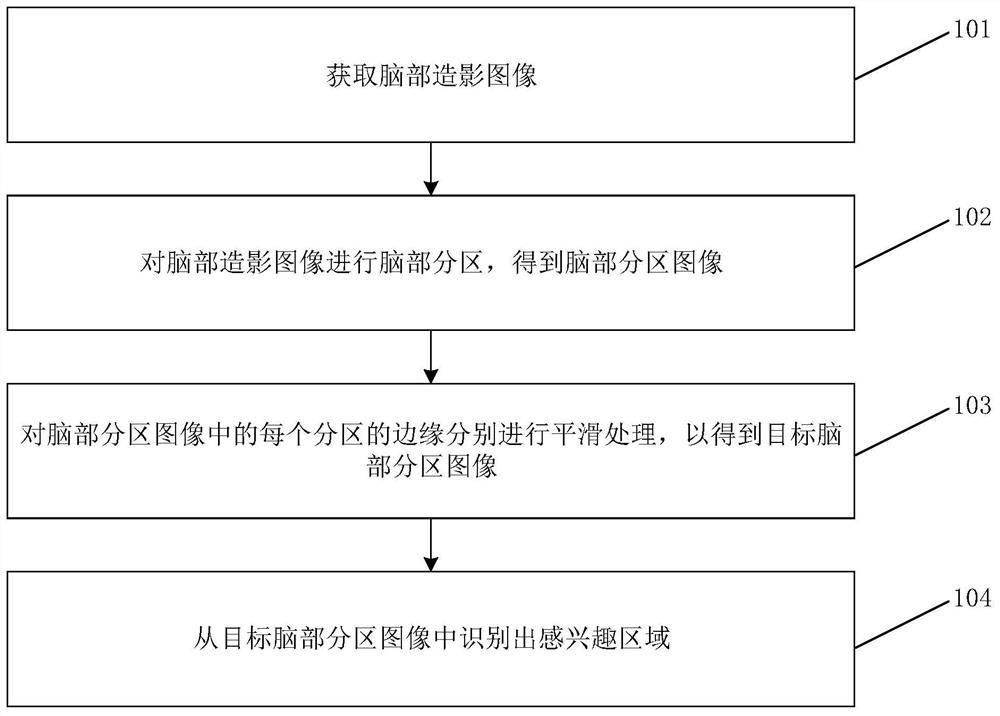
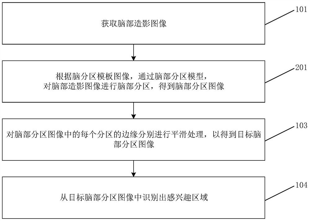
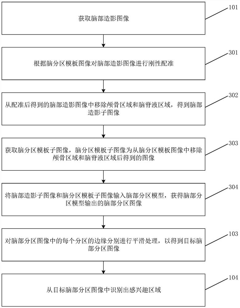
[0069] Specific embodiments of the present disclosure will be described in detail below in conjunction with the accompanying drawings. It should be understood that the specific embodiments described here are only used to illustrate and explain the present disclosure, and are not intended to limit the present disclosure.

[0070] figure 1 It is a flow chart of a brain imaging image processing method according to an exemplary embodiment of the present disclosure. like figure 1 As shown, the method includes steps 101 to 104.

[0071] In step 101, brain imaging images are acquired. The brain imaging image can be a three-dimensional imaging image for the brain obtained by any means, that is, a volume image, for example, it can be a dicom format volume image obtained by CT angiography (CTA, CTangiography), or it can be Dicom format volume images obtained by MRA magnetic resonance angiography (magneticresonance angiography, MRA), etc.

[0072] Wherein, after the brain imaging im...


PUM


Abstract

The invention relates to a brain contrast image processing method and device, a medium and electronic equipment. The method comprises the steps that a brain contrast image is acquired; brain partitionis performed on the brain contrast image to obtain a brain partition image; the edge of each partition in the brain partition image is smoothed to obtain a target brain partition image; and a regionof interest is identified from the target brain partition image. Thus, the acquired cerebral angiography image can be partitioned at first; then region-of-interest identification is respectively carried out on the subarea images; the whole processing method can be automatically executed, manual operation is not needed, a manual method does not need to be adopted for visual evaluation, the recognition difficulty and cost of the region of interest are reduced. The problems that due to visual evaluation conducted through the manual method, consumed time is long, efficiency is low, recognition results are inconsistent, and consequently the reliability of judgment results is affected are solved.

Description

technical field [0001] The present disclosure relates to the field of computers, and in particular, to a method, device, medium and electronic equipment for processing brain imaging images. Background technique [0002] Neuroimaging plays an important clinical role in assessing the severity and prognosis of ischemic stroke and guiding clinical treatment. Within hours of the onset of acute ischemic stroke, local low-density shadows may appear on brain non-contrast CT (NCCT, ie plain CT) scan images. This hypodensity is secondary to changes in water content due to focal cerebral ischemia. This early ischemic change represents cytotoxic edema of brain tissue, or irreversible damage. In order to accurately evaluate early ischemic changes, Barber et al. developed the Alberta Stroke Program Early CT Score (ASPECTS), which is a scale of cerebral ischemia based on NCCT images, and aims to measure the degree of cerebral infarction in acute stroke. Extent and distribution of early ...

Claims


Application Information

Patent Timeline
IPC IPC(8): G06T7/11G06K9/32G06T5/00
CPCG06T7/11G06T5/003G06T2207/10081G06T2207/20192G06T2207/30016G06V10/25
Inventor 杨俊冯莹莹马丽娟
Owner 沈阳东软智能医疗科技研究院有限公司
Why Eureka
Try Eureka