Three-dimensional image splicing method, device, equipment and system and storage medium

A three-dimensional image and medical imaging system technology, applied in the field of three-dimensional image stitching methods, systems and storage media, equipment, and devices, can solve the problems of prolonging scanning time, affecting the quality of three-dimensional tomographic images, and reducing the spatial resolution of scanned images.

Pending Publication Date: 2020-11-27
SHANGHAI UNITED IMAGING HEALTHCARE
View PDF9 Cites 8 Cited by
  • Summary
  • Abstract
  • Description
  • Claims
  • Application Information

AI Technical Summary

Problems solved by technology

Due to the motion artifact brought by mechanical movement and the time delay caused by the thermal electron emission mechanism, the spatial resolution of the scanned image is reduced and the scanning time is prolonged. Motion artifacts are easily generated during the shooting process, thus affecting the three-dimensional tomographic image. the quality of

Method used


Image

  • Three-dimensional image splicing method, device, equipment and system and storage medium
  • Three-dimensional image splicing method, device, equipment and system and storage medium
  • Three-dimensional image splicing method, device, equipment and system and storage medium

Examples


Embodiment Construction

[0063] In order to make the purpose, technical solution and advantages of the present application clearer, the present application will be further described in detail below in conjunction with the accompanying drawings and embodiments. It should be understood that the specific embodiments described here are only used to explain the present application, and are not intended to limit the present application.

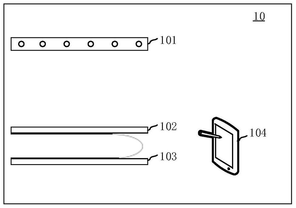
[0064] The three-dimensional image stitching method provided in the embodiment of the present application can be applied to figure 1 The X-ray medical imaging system 10 shown includes an array X-ray source 101 , a compression plate 102 , a detector 103 and a computer device 104 .

[0065] Wherein, the array X-ray source 101 is used to emit X-rays. The array X-ray source includes a plurality of X-ray sources, and each X-ray source is a field emission X-ray source and can emit X-rays. Optionally, the array X-ray source includes one or more of a linear array ray source and ...


PUM


Abstract

The invention relates to a three-dimensional image splicing method, device, equipment and system and a storage medium. The method is applied to an X-ray medical imaging system, the X-ray medical imaging system comprises a detector and array X-ray sources, the array X-ray sources comprise a plurality of X-ray sources with different projection angles, and imaging areas corresponding to the X-ray sources are formed on the detector. The method comprises the following steps: acquiring projection data of each region of a to-be-detected part, wherein the projection data is generated by exposing eachregion by X-ray sources which are located at at least two different projection angles relative to the regions among the array X-ray sources; respectively carrying out three-dimensional image reconstruction on the projection data of each region to obtain reconstructed image blocks; and splicing the reconstructed image blocks according to the relative position relationship among the reconstructed image blocks so as to obtain a three-dimensional reconstructed image of the to-be-detected part. By adopting the method, the quality of the three-dimensional reconstructed image can be improved.

Description

technical field [0001] The present application relates to the technical field of image processing, in particular to a three-dimensional image mosaic method, device, equipment, system and storage medium. Background technique [0002] With the continuous development of X-ray technology, most of the X-ray data of the patient are collected by the X-ray machine when breast examination is performed on the patient, and then the image reconstruction is performed on the collected data to obtain the medical image of the patient's breast area, and then the medical After analyzing the image, the patient's image analysis result can be obtained. [0003] Generally, in traditional mammary X-ray medical imaging products, a single light source with hot cathode rotation is usually used. In order to perform multi-view X-ray scanning, the X-ray light source is fixed on the rotating frame and performs arc motion for X-ray scanning. Due to the motion artifact caused by mechanical movement and th...

Claims


Application Information

Patent Timeline
Patent Type & Authority Applications(China)
IPC IPC(8): A61B6/00A61B6/03G06T3/40G06T17/00
CPCA61B6/032A61B6/035A61B6/4014A61B6/4266A61B6/4429A61B6/5241A61B6/5258G06T17/00G06T3/4038G06T2200/32
Inventor 牛杰张宇
Owner SHANGHAI UNITED IMAGING HEALTHCARE
Why Eureka
Try Eureka