Ultrasonic elastography cornea detection method, device and system and storage medium

An ultrasonic elastography and elastography technology, applied in ultrasonic/sonic/infrasonic diagnosis, eye examination, sonic diagnosis, etc., can solve problems such as cumbersome operations, corneal damage, and inability to apply clinical diagnosis to reduce injuries and improve accuracy Effect

Active Publication Date: 2020-12-04
SHENZHEN UNIV
View PDF7 Cites 6 Cited by
  • Summary
  • Abstract
  • Description
  • Claims
  • Application Information

AI Technical Summary

Problems solved by technology

The disadvantage of this method is that the excitation acquisition step needs to be performed three times, and the operation is cumbersome
In the existing measurement schemes, the measurement of the biomechanical properties of the cornea is obtained through in vitro experiments. The in vitro corneal measurement experiments can include axial mechanical stretching experiments and expansion experiments, but these two methods will cause damage to the cornea. Destruction of reversal, can not be applied to clinical diagnosis
A method for clinical diagnosis of corneal biomechanical properties is currently lacking

Method used


Image

  • Ultrasonic elastography cornea detection method, device and system and storage medium
  • Ultrasonic elastography cornea detection method, device and system and storage medium
  • Ultrasonic elastography cornea detection method, device and system and storage medium

Examples


Embodiment 1

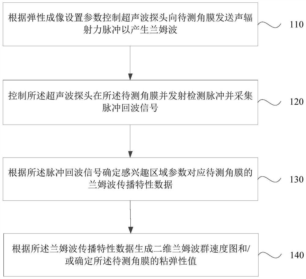
[0034] figure 1 This is a flow chart of an ultrasonic elastography corneal detection method provided in the first embodiment of the present invention. This embodiment can be applied to the situation of detecting the biomechanical properties of the cornea of ​​the eye in a clinical state, and the method can be performed by an ultrasonic elastography corneal device. , the device can be implemented by means of hardware and / or software, see figure 1 , the method provided by the embodiment of the present invention specifically includes the following steps:

[0035] Step 110: Control the ultrasonic probe to send acoustic radiation force pulses to the cornea to be measured according to the elastography setting parameters to generate Lamb waves, wherein the ultrasonic probe includes at least two channel array elements.

[0036] The elastography setting parameters may be parameters for setting the ultrasonic probe, and the elastography setting parameters may include information such a...

Embodiment 2

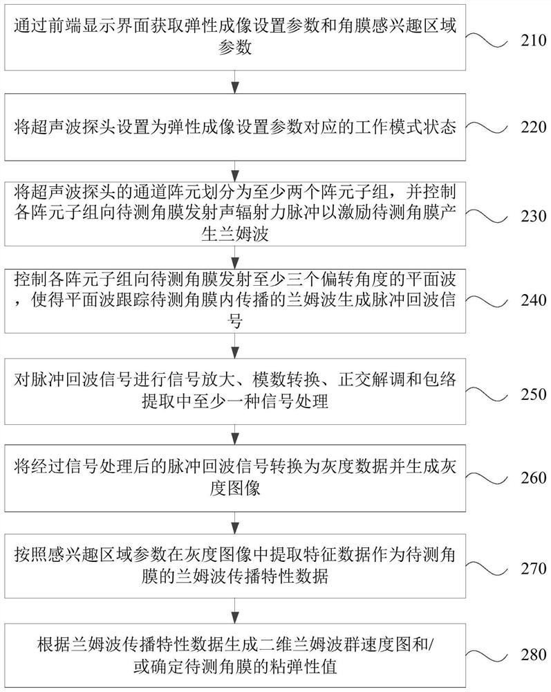
[0051] figure 2 It is a flow chart of a method for ultrasonic elastography cornea detection provided by Embodiment 2 of the present invention. The embodiment of the present invention is based on the embodiment of the above invention. See figure 2 , the method provided in the embodiment of the present invention specifically includes the following steps:

[0052] Step 210, obtaining elastography setting parameters and corneal region-of-interest parameters through the front-end display interface.

[0053] Wherein, the front-end input display interface may be a display module for human-computer interaction with the user, and the front-end input display interface may receive user input information and display specific data of the cornea to be measured.

[0054] Specifically, the user can operate the front-end display interface, and input elastography setting parameters and corneal region-of-interest parameters to the front-end by inputting information, dragging and dragging the ...

Embodiment 3

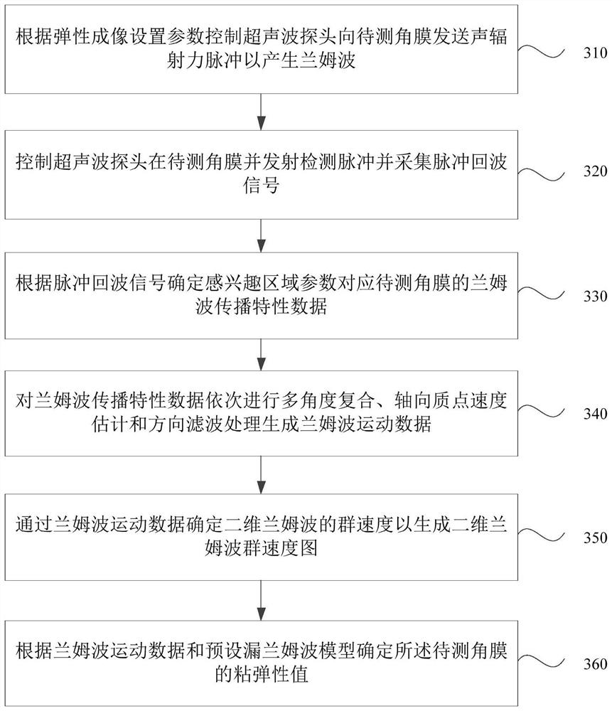
[0078] image 3 It is a flow chart of an ultrasonic elastography corneal detection method provided in Embodiment 3 of the present invention, see image 3 , on the basis of the above application embodiment, the method provided by the embodiment of the present invention includes the following steps:

[0079] Step 310: Control the ultrasonic probe to send acoustic radiation force pulses to the cornea to generate Lamb waves according to elastography setting parameters, wherein the ultrasonic probe includes at least two channel elements.

[0080] Step 320 , controlling the ultrasonic probe to be placed on the cornea to be tested to transmit detection pulses and collect pulse echo signals.

[0081] Step 330: Determine the lamb wave propagation characteristic data corresponding to the parameters of the region of interest corresponding to the cornea to be measured according to the pulse echo signal.

[0082] Step 340 , sequentially perform multi-angle compounding, axial particle vel...


PUM


Abstract

The invention discloses an ultrasonic elastography cornea detection method, device and system and a storage medium. The method comprises the steps of controlling an ultrasonic probe to send an acoustic radiation force pulse to a to-be-detected cornea according to an elastography setting parameter, so as to generate a lamb wave, wherein the ultrasonic probe at least comprises two channel array elements; controlling the ultrasonic probe to be in the cornea to be detected, transmitting a detection pulse and acquiring a pulse echo signal; determining lamb wave propagation characteristic data of the cornea to be measured corresponding to the region-of-interest parameter according to the pulse echo signal; and generating a two-dimensional lamb wave group velocity diagram and / or determining the viscoelasticity value of the cornea to be measured according to the lamb wave propagation characteristic data. According to the embodiment of the invention, the ultrasonic probes of the multichannel array elements excite the cornea to obtain the propagation characteristics of the lamb waves, and the biomechanical characteristics of the cornea are determined according to the propagation characteristics of the lamb waves in the cornea, so that the clinical detection of the cornea is realized, the resolution of detection imaging is improved, and the accuracy of a detection result is enhanced.

Description

technical field [0001] Embodiments of the present invention relate to the technical field of computer applications, and in particular, to an ultrasonic elastography cornea detection method, device, system and storage medium. Background technique [0002] The incidence of keratoconus in the population is 5 in 1,000 and usually results in highly irregular astigmatism and varying degrees of visual impairment. When keratoconus occurs, the corneal tissue will gradually thin, and its shape will gradually change from a normal circle to a cone, and the front end of the corneal tissue will be more prominent, forming a cone. Keratoconus is present in all ethnic groups and affects both men and women equally. The onset of primary keratoconus is usually early in adulthood, and the disease progresses for 10-20 years. The incidence of keratoconus is mainly in the career rise and child-rearing period of patients, so the impact of keratoconus on the life of patients may be much more seriou...

Claims


Application Information

Patent Timeline
Patent Type & Authority Applications(China)
IPC IPC(8): A61B8/10A61B3/107A61B3/10
CPCA61B3/107A61B3/1005A61B8/10A61B8/485A61B8/5207Y02T90/00
Inventor 张新宇赵林风陈昕陈思平
Owner SHENZHEN UNIV
Why Eureka
Try Eureka