CT arbitrary section ultrasonic visual field simulation auxiliary ablation path planning method and system

A technology of path planning and ultrasound, which is applied in the field of medical image processing, can solve the problems of complex volume ultrasound acquisition process, disjointed and difficult acquisition of the actual implementation process of surgical planning, and achieve the effect of improving implementability and accuracy

Active Publication Date: 2020-12-08
THE FIFTH MEDICAL CENT OF CHINESE PLA GENERAL HOSPITAL
View PDF14 Cites 2 Cited by
  • Summary
  • Abstract
  • Description
  • Claims
  • Application Information

AI Technical Summary

Problems solved by technology

However, the preoperative planning based on CT images often deviates greatly from the intraoperative puncture path guided by ultrasound, which has become one of the challenges of precise ablation.
[0004] Three-dimensional space display is the advantage of three-dimensional space needle insertion planning, but it also has great limitations. Specifically, there is a large difference between the CT observation field of view and the intraoperative real-time ultrasound field of view, resulting in path planning only based on CT images and actual ultrasound guidance. The maneuverability of lower needle insertion does not match, which eventually leads to a disconnect between surgical planning and actua

Method used


Image

  • CT arbitrary section ultrasonic visual field simulation auxiliary ablation path planning method and system
  • CT arbitrary section ultrasonic visual field simulation auxiliary ablation path planning method and system
  • CT arbitrary section ultrasonic visual field simulation auxiliary ablation path planning method and system

Examples


Embodiment Construction

[0023] Image-guided minimally invasive technology has become one of the hotspots in tumor treatment. In order to achieve precise inactivation of tumors under the guidance of ultrasound images, three-dimensional visualization preoperative planning is an essential means, but preoperative planning based on CT images is often different from ultrasound-guided Therefore, the invention of CT three-dimensional images with ultrasound field of view is the key method to solve this problem.

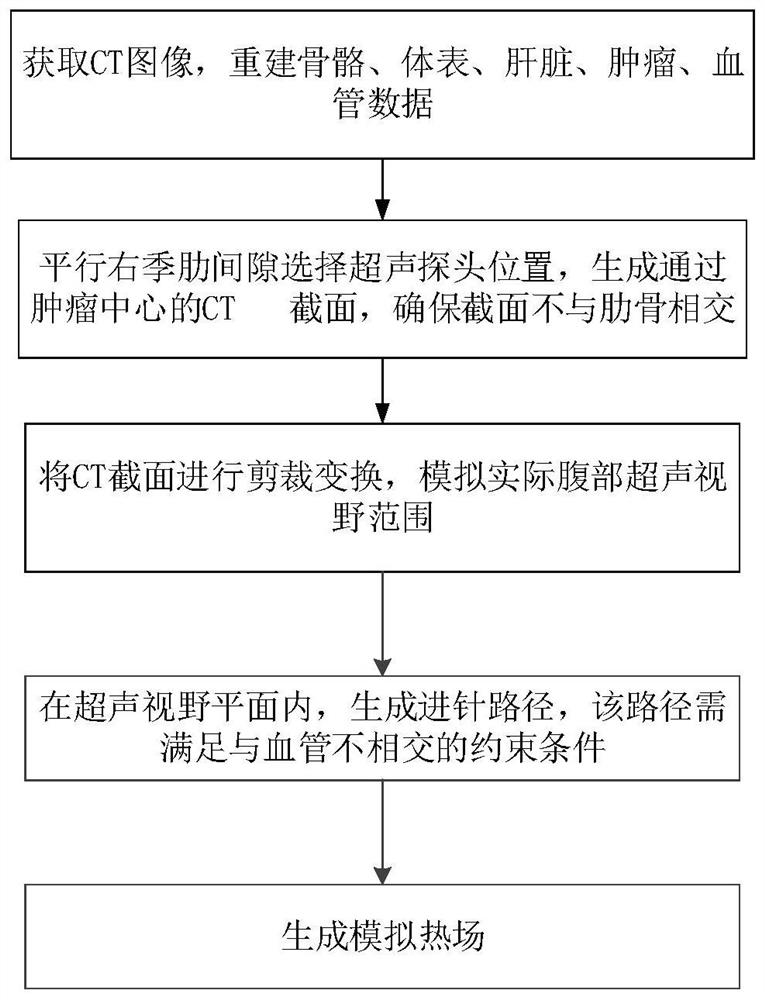
[0024] Such as figure 1 As shown, this CT ablation path planning method assisted by ultrasonic field of view simulation in any slice, includes the following steps:

[0025] (1) Obtain CT images and reconstruct bone, body surface, liver, tumor, and blood vessel data;

[0026] (2) Select the position of the ultrasound probe parallel to the right intercostal space, and generate a CT section through the tumor center to ensure that the section does not intersect with the ribs;

[0027] (3) Cutting and t...


PUM


Abstract

The invention discloses a CT arbitrary section ultrasonic visual field simulation auxiliary ablation path planning method and system. The method comprises the steps of : (1) obtaining a CT image, andreconstructing data of bone, body surface, liver, tumor and blood vessel; (2) selecting the position of an ultrasonic probe parallel to a right hypochondrium gap, generating a CT section passing through a tumor center, and ensuring that the section does not intersect with a rib; (3) carrying out clipping transformation on the CT section, and simulating an actual abdominal ultrasonic visual field range; (4) generating a needle inserting path in the ultrasonic visual field plane, and the path should meet the constraint condition that the path does not intersect with the blood vessel; and (5) generating a simulated thermal field; judging the space coverage relation between the thermal field and the tumor, and if the space coverage relation is not complete, obtaining the parallel cross sectionof a plane along the long-axis direction of the coronary tumor, and executing the step (3) until the thermal field completely covers the tumor.

Description

technical field [0001] The present invention relates to the technical field of medical image processing, and in particular to a method for assisting ablation path planning with ultrasound field of view simulation at any slice of CT, and a system for planning an ablation path with the assistance of ultrasound field of view simulation at any slice of CT. Background technique [0002] Ultrasound-guided microwave, radiofrequency and other ablation therapy is a new local treatment method for liver cancer compared with surgery. This technology converts electromagnetic waves into heat energy through microwave or radiofrequency needles guided by ultrasound, and transmits the energy to the tumor area to achieve tumor localization. Complete coagulation necrosis has been recommended by NCCN, BCLC and other international guidelines as the first-line treatment for small liver cancer. However, ultrasound-guided ablation of liver cancer is a comprehensive process involving various knowledg...

Claims


Application Information

Patent Timeline
IPC IPC(8): A61B34/00A61B34/20A61B18/12
CPCA61B34/00A61B34/20A61B18/12A61B2034/107A61B2034/101A61B2034/105A61B2034/108A61B2034/2063A61B2034/2065A61B2018/00577
Inventor 梁萍于杰董立男程志刚刘方义韩治宇
Owner THE FIFTH MEDICAL CENT OF CHINESE PLA GENERAL HOSPITAL
Why Eureka
Try Eureka