Retinal fundus vessel segmentation method based on deep multi-scale attention convolutional neural network

A convolutional neural network and attention technology, applied in retinal vessel segmentation, computer technology and pattern recognition, to achieve clear segmentation probability map, accurate background segmentation, and avoid feature loss

Active Publication Date: 2020-12-18
BEIHANG UNIV
View PDF4 Cites 40 Cited by
  • Summary
  • Abstract
  • Description
  • Claims
  • Application Information

AI Technical Summary

Problems solved by technology

[0006] In order to solve the problems existing in the existing image segmentation technology, the present invention provides a retinal fundus blood vessel segmentation method based on deep multi-scale attention convolutional neural network

Method used


Image

  • Retinal fundus vessel segmentation method based on deep multi-scale attention convolutional neural network
  • Retinal fundus vessel segmentation method based on deep multi-scale attention convolutional neural network
  • Retinal fundus vessel segmentation method based on deep multi-scale attention convolutional neural network

Examples


Embodiment Construction

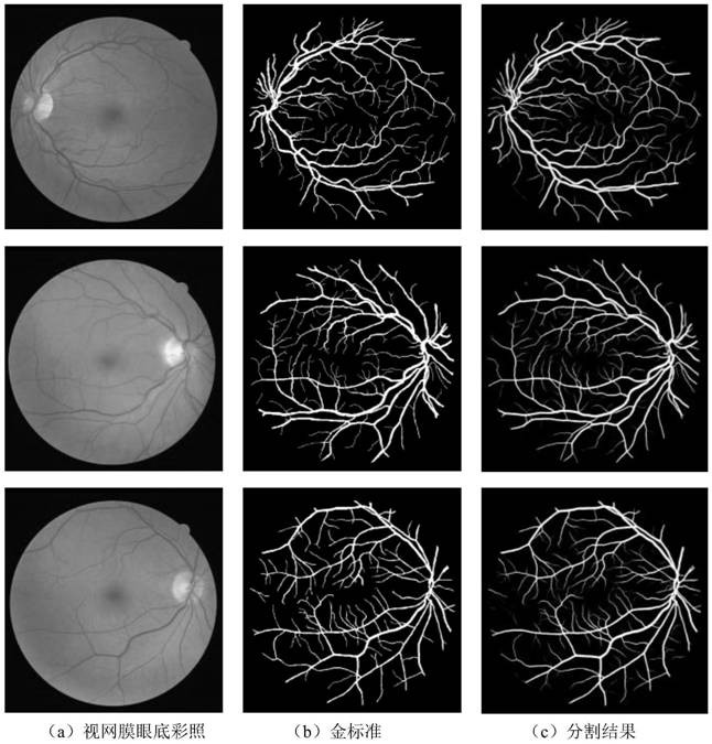
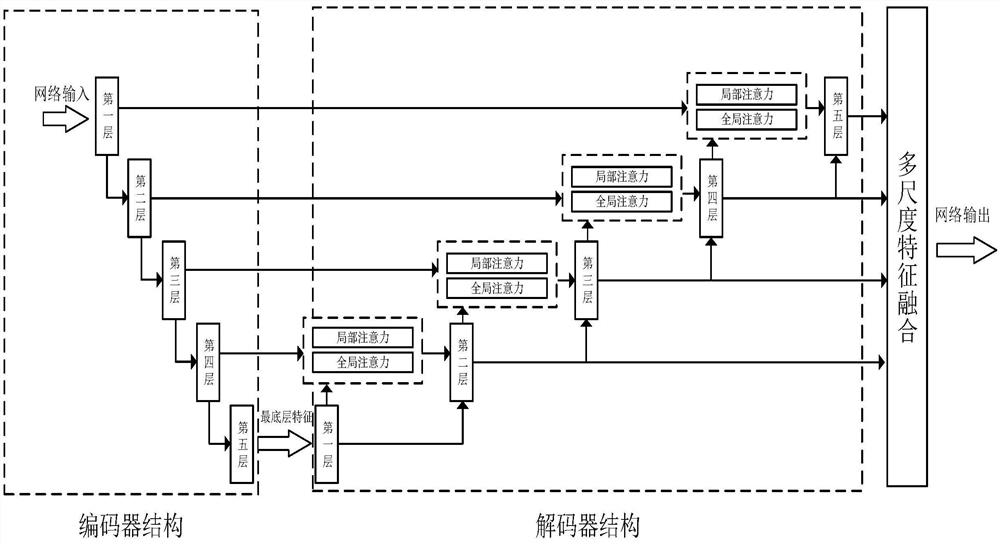
[0023] According to one embodiment of the present invention, a retinal fundus vessel segmentation method based on a deep multi-scale attentional convolutional neural network is proposed. A dual attention mechanism is introduced in the connection path between the encoder and the decoder of the U-Net architecture, and a multi-scale feature fusion module is introduced at the output of each layer of the decoder to finally obtain the retinal fundus blood vessel segmentation result.

[0024] Below in conjunction with the accompanying drawings, the specific implementation of a retinal fundus blood vessel segmentation method based on a deep multi-scale attention convolutional neural network proposed by the present invention will be described in detail:

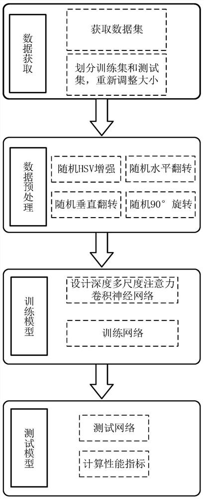
[0025] Step 1: Obtain DRIVE, an internationally publicized color retinal fundus blood vessel dataset;

[0026] Step 2: Select the pictures used for training in the data set, adjust their size to 512×512 pixels, design a random data en...


PUM


Abstract

The invention provides a retinal fundus vessel segmentation method based on a deep multi-scale attention convolutional neural network. An internationally disclosed retinal fundus vessel data set DRIVEis adopted to perform validity verification: firstly, dividing the retinal fundus vessel data set DRIVE into a training set and a test set, and adjusting the picture size to 512*512 pixels; then, enabling the training set to be subjected to four random preprocessing links to achieve a data enhancement effect; designing a model structure of the deep multi-scale attention convolutional neural network, and inputting the processed training set into the model for training; and finally, inputting the test set into the trained network, and testing the model performance. The main innovation point ofthe method is that a double attention module is designed, so that the whole model pays more attention to segmentation of small blood vessels; and a multi-scale feature fusion module is designed, so that the global feature extraction capability of the whole model on the segmented image is stronger. The segmentation accuracy of the model on a DRIVE data set is 96.87%, the sensitivity is 79.45%, thespecificity is 98.57, and the method is superior to classical UNet and an existing most advanced segmentation method.

Description

technical field [0001] The present invention provides a retinal vessel segmentation method based on a deep multi-scale attentional convolutional neural network. It provides a new method for the application of computer technology in the field of retinal blood vessel segmentation, and belongs to the field of computer technology and pattern recognition. Background technique [0002] In the medical field, retinal fundus image analysis is an important means for doctors to screen fundus diseases (such as age-related macular degeneration, glaucoma, diabetic retinopathy) and some cardiovascular diseases (such as hypertension). The symptoms of these diseases will become more obvious as the patient grows older. When the eye diseases are severe, they may even lead to blindness. If they are not treated in time, they may lead to death. Therefore, a fast and accurate intelligent retinal fundus image analysis method is one of the core technologies urgently needed in the field of ophthalmo...

Claims


Application Information

Patent Timeline
Patent Type & Authority Applications(China)
IPC IPC(8): G06T7/00G06N3/08G06N3/04G06K9/62
CPCG06T7/0012G06N3/08G06T2207/30041G06N3/045G06F18/214
Inventor 李阳张越
Owner BEIHANG UNIV
Why Eureka
Try Eureka