
Method and system for determining cancer therapy effects via imaging omics features

A radiomics and response technology, applied in informatics, image enhancement, image analysis, etc., can solve the problem of inability to solve the risk prediction of tumor recurrence and metastasis, and achieve low radiation damage risk, high repeatability, and good radiotherapy efficacy. Effect

Pending Publication Date: 2021-01-22
BEIJING CANCER HOSPITAL PEKING UNIV CANCER HOSPITAL
View PDF5 Cites 2 Cited by
  • Summary
  • Abstract
  • Description
  • Claims
  • Application Information

AI Technical Summary

Problems solved by technology

[0004] The main purpose of the present invention is to overcome the defects of the above-mentioned prior art, and provide a method and system for judging cancer treatment response through radiomics features, so as to solve the problem of tumor recurrence and metastasis risk prediction and the risk of radiation damage that cannot be solved by the prior art. prediction problem

Method used


Image

  • Method and system for determining cancer therapy effects via imaging omics features
  • Method and system for determining cancer therapy effects via imaging omics features
  • Method and system for determining cancer therapy effects via imaging omics features

Examples


Embodiment 1

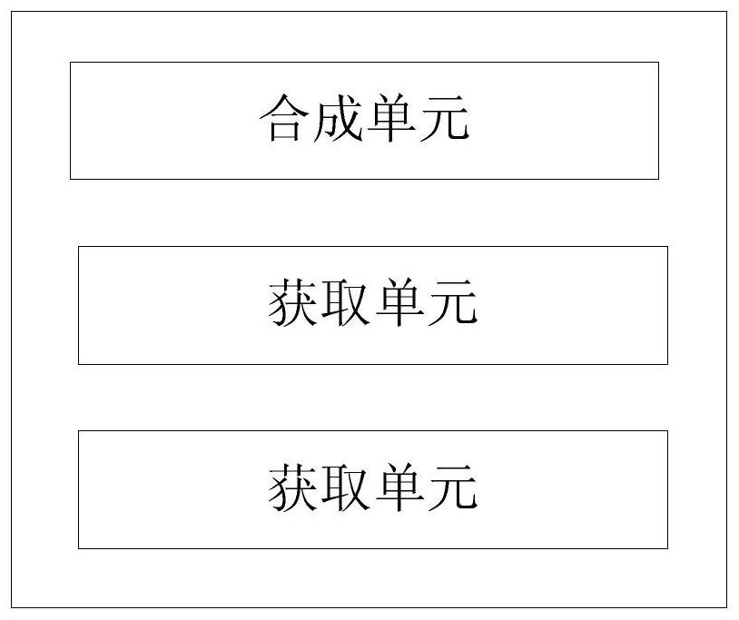
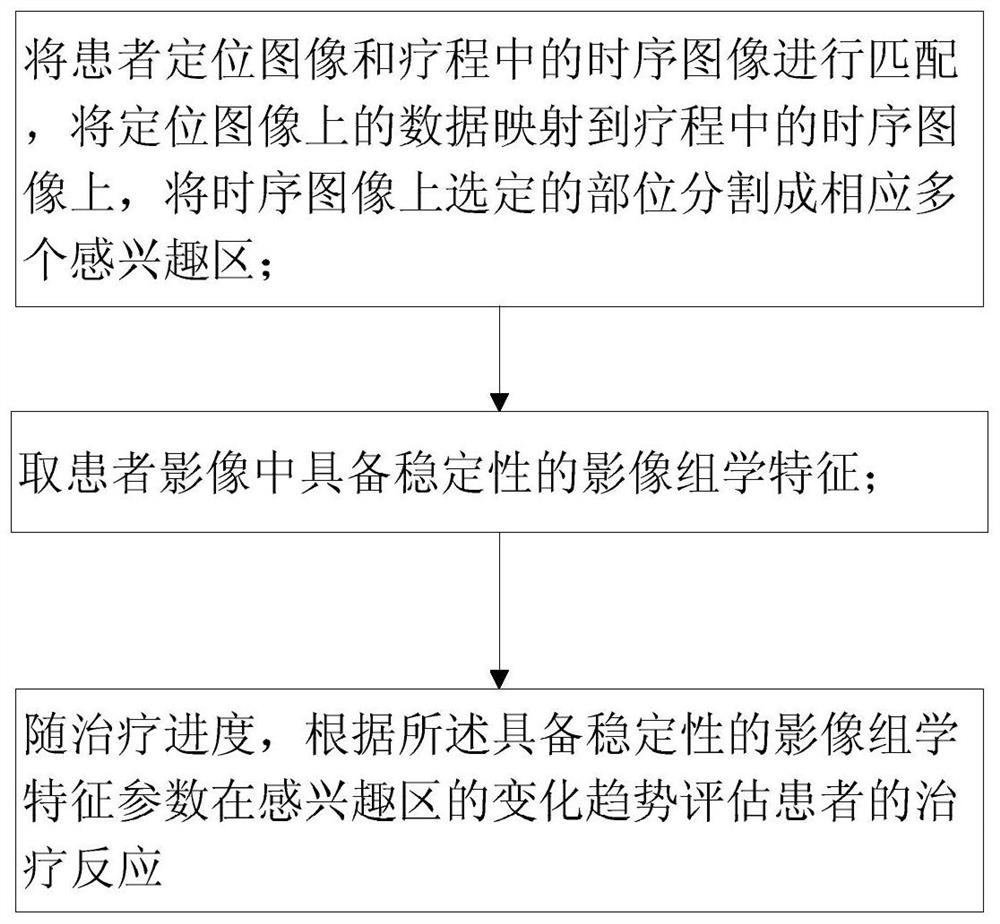
[0036] A system for judging cancer treatment response by radiomics features, such as figure 1 As shown, it includes: a synthesizing unit, which is used to match the positioning image of the patient with the time-series images in the course of treatment, map the data on the positioning image to the time-series images in the course of treatment, and divide the selected parts on the time-series images into corresponding multiple parts. A region of interest; the acquisition unit is used to extract radiomics features with stability in patient images; the analysis unit is used to evaluate the treatment response of the patient according to the change trend of the radiomics characteristic parameters in the region of interest as the treatment progresses .

[0037] Time-series images include time-series CBCT images, or other imaging with similar functions. The advantage of time-series CBCT image series is that it can provide multi-dimensional correlation information such as changes in r...

Embodiment 2

[0046] The radiomics features with stability include radiomics features with time stability; the radiomics features with time stability refer to images in images collected at the same location under the same conditions at different times The omics features are consistent and are time-stable radiomics features.

[0047] At the same time avoid the loss of important original information, and use the consistency correlation coefficient (CCC) to evaluate the time stability and cross-modal equivalence of radiomics features. When the consistency correlation coefficient (CCC) is greater than a certain value, the image The omics characteristics are consistent.

[0048] In order to evaluate and screen stable CBCT radiomics features, the present invention optimizes the preprocessing method. In one embodiment, Pyradiomics software is used to realize the automatic calculation of radiomics features, and two CBCT images collected at different times based on the same conditions are used. Rad...

Embodiment 3

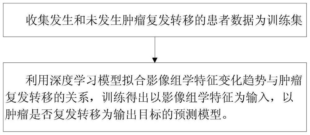
[0062] The method of radiation injury risk analysis and model establishment is the same as that of Embodiment 2, the difference being that the composition of the training set is different.


PUM


Abstract

Disclosed is a method and system for determining cancer therapy effects via imaging omics features, including a synthesis unit which is used for matching the patient positioning image with the timingimages in the treating process, wherein the data on the positioning image is mapped to the timing images in the treating process; the selected part on the timing image is cut into multiple corresponding area of interest; an acquisition unit used for extrating imaging omics features with stability on the patient image; and an analysis unit used for assessing the treating effects of the patients based on the changes of the imaging omics feature parameters in the areas of interest along with the treatment progress. The scheme has the following advantages of high stability and repeatability; the timing image optic features changes sharply along with the treatment gradation and accumulated dose; the early individuation prediction of cancer recurrence and metastasis or radiation damage to the normal tissue are obtained, so the control probability of the cancer is improved while the radiation damage to the normal tissue and cancer relapse are reduced.

Description

technical field [0001] The present invention relates to the field of radiotherapy equipment, in particular to a method and system for judging cancer treatment response through radiomics features. Background technique [0002] Radiotherapy is one of the main treatment methods for cancer, and there is a radiation dosimetric contradiction between tumor control and organ damage. The radiotherapy effect of cancer is positively correlated with the radiation dose of the target area, but the increased radiation damage, such as the risk of pneumonia (lung exposure) in lung cancer, is also a key constraint factor for further increasing the dose of the target area, and it is also the main reason for the interruption or even failure of radiotherapy one of the reasons. Putting the lungs is an inflammatory reaction after normal lung tissue is damaged by radiation. The main clinical manifestation is diffuse alveolar damage, which may develop into radiation-induced pulmonary fibrosis in se...

Claims


Application Information

Patent Timeline
Patent Type & Authority Applications(China)
IPC IPC(8): G16H50/20G06T7/11G06T7/30
CPCG16H50/20G06T7/11G06T7/30G06T2207/10081G06T2207/10088G06T2207/10104G06T2207/30096G06T2207/20081G06T2207/30061
Inventor 张艺宝黄宇亮李晨光吴昊刘宏嘉
Owner BEIJING CANCER HOSPITAL PEKING UNIV CANCER HOSPITAL
Learn More