Lung image processing method and device, electronic equipment and storage medium

An image processing and lung technology, applied in the field of medical image processing, can solve the problems of high medical quality requirements, missed detection of tuberculosis, and low screening efficiency, so as to improve screening efficiency, reduce manual participation, and solve missed tuberculosis The effect of detecting phenomena

Inactive Publication Date: 2021-03-02
江西中科九峰智慧医疗科技有限公司
View PDF6 Cites 0 Cited by
  • Summary
  • Abstract
  • Description
  • Claims
  • Application Information

AI Technical Summary

Problems solved by technology

[0003] In the screening process of pulmonary tuberculosis, usually only the obvious lesions in the frontal chest X-ray images can be screened, but the tiny lesions in the frontal chest X-ray images cannot be screened, and tuberculosis is prone to missed detection.
At the same time, the screening program has extremely high requirements on the medical quality of doctors, and the corresponding screening operations need to be completed manually by doctors, and the overall screening efficiency is not high.

Method used


Image

  • Lung image processing method and device, electronic equipment and storage medium
  • Lung image processing method and device, electronic equipment and storage medium
  • Lung image processing method and device, electronic equipment and storage medium

Examples


Embodiment 1

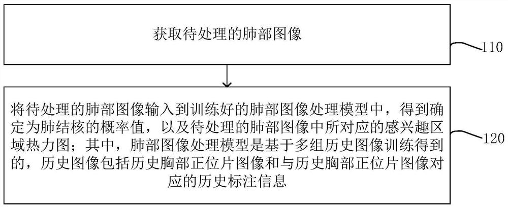
[0034] figure 1 It is a flow chart of a lung image processing method provided in Embodiment 1 of the present invention. This embodiment is applicable to processing lung images and screening for tuberculosis. The method can be executed by a lung image processing device. The lung image processing device can be implemented by software and / or hardware, and the lung image processing device can be configured on an electronic computing device, specifically including the following steps:

[0035] S110. Acquire a lung image to be processed.

[0036] Exemplarily, the image of the lungs to be processed may be an image to be identified whether there are tuberculosis lesions in the image.

[0037] Optionally, the image of the lungs to be processed may be an anteroposterior view of the chest.

[0038] S120. Input the lung image to be processed into the trained lung image processing model to obtain the probability value determined as pulmonary tuberculosis and the thermal map of the region...

Embodiment 2

[0055] Image 6 It is a flow chart of the lung image processing method provided by Embodiment 2 of the present invention. The embodiment of the present invention may be combined with various alternative solutions in the foregoing embodiments. In the embodiment of the present invention, optionally, the training method of the lung image processing model includes: acquiring multiple sets of historical chest front view images and historical annotation information corresponding to the historical chest front view images, wherein the history The label information is the information after labeling the pulmonary tuberculosis in the historical chest front view images; input multiple groups of historical chest front view images and the historical label information corresponding to the historical chest front view images into the lung image processing model to be trained In , iteratively trains the lung image processing model to be trained to obtain a trained lung image processing model. ...

Embodiment 3

[0103] Figure 13 It is a flow chart of the lung image processing method provided in Embodiment 3 of the present invention. The embodiment of the present invention may be combined with various alternative solutions in the foregoing embodiments. In the embodiment of the present invention, optionally, before inputting multiple sets of historical chest front view images and historical annotation information corresponding to the historical chest front view images into the lung image processing model to be trained, the The method also includes: for any set of acquired historical chest front view images and historical annotation information corresponding to the historical chest front view images, the following steps are performed: preprocessing the historical chest front view images and the historical annotation information to obtain Target historical chest front view image and target history annotation information; correspondingly, input multiple groups of history chest front view ...


PUM


Abstract

The embodiment of the invention discloses a lung image processing method and device, electronic equipment and a storage medium. The method comprises the following steps: acquiring a lung image to be processed; and inputting the lung image to be processed into a trained lung image processing model to obtain a probability value determined as pulmonary tuberculosis and a corresponding region-of-interest thermodynamic diagram in the lung image to be processed, wherein the lung image processing is obtained on the basis of training of multiple groups of historical images, and the historical images comprise historical chest orthographic film images and historical labeling information corresponding to the historical chest orthographic film images. Therefore, the pulmonary tuberculosis missing detection phenomenon in the pulmonary tuberculosis screening process is improved, the artificial participation degree in the pulmonary tuberculosis screening process is reduced, the pulmonary tuberculosisscreening efficiency is improved, and doctors are assisted in completing pulmonary tuberculosis diagnosis.

Description

technical field [0001] Embodiments of the present invention relate to medical image processing technologies, and in particular, to a lung image processing method, device, electronic equipment, and storage medium. Background technique [0002] In the medical field, tuberculosis is currently one of the infectious diseases with the highest fatality rate. At present, doctors use the naked eye to locate and identify tuberculosis-related lesions in the target orthotopic chest radiograph image to complete the tuberculosis screening operation. [0003] In the screening process of pulmonary tuberculosis, usually only the obvious lesions in the frontal chest radiograph can be screened, but the tiny lesions in the frontal chest radiograph cannot be screened, and tuberculosis is prone to missed detection. At the same time, the screening program has extremely high requirements on the medical quality of doctors, and the corresponding screening operations need to be completed manually by d...

Claims


Application Information

Patent Timeline
Patent Type & Authority Applications(China)
IPC IPC(8): G06T7/00G06T7/143G16H30/40G16H50/20
CPCG06T7/0012G06T2207/30061G06T2207/30096G06T7/143G16H30/40G16H50/20
Inventor 马勇李祎吴文辉
Owner 江西中科九峰智慧医疗科技有限公司
Why Eureka
Try Eureka