Shear wave elastic imaging method and system
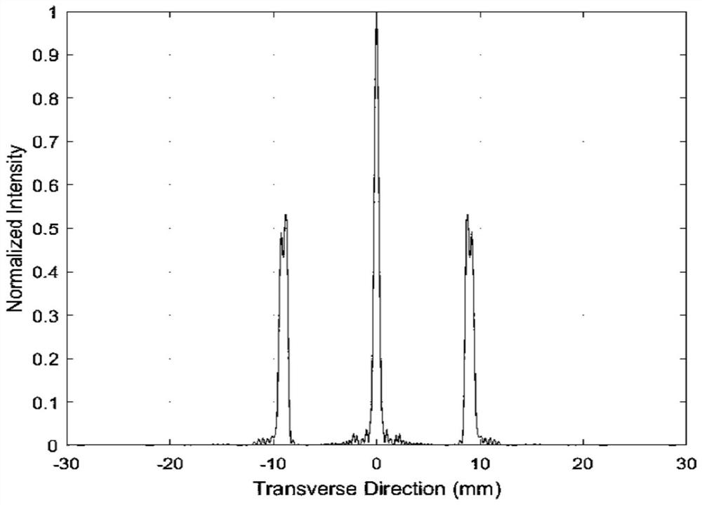
An elastic imaging and shear wave technology, applied in the field of ultrasonic scanning, can solve the problem of uneven intensity distribution and achieve the effect of uniform intensity distribution
- Summary
- Abstract
- Description
- Claims
- Application Information
AI Technical Summary
Problems solved by technology
Method used
Image
Examples
Embodiment 1
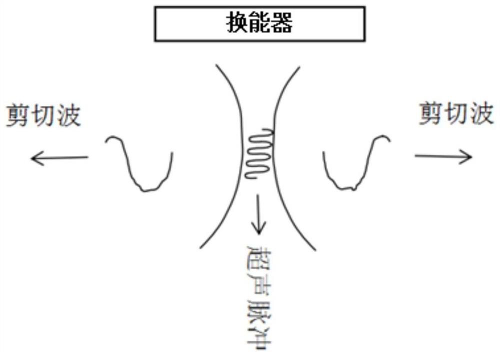
[0055] Acoustic waves propagate through tissue much faster (approximately 1000 times) than shear waves, so the propagation of shear waves in tissue in the transverse direction can be fully tracked. By measuring the shear wave velocity in the region of interest, it is possible to provide Two-dimensional quantitative elastograms of tissue. A very high frame rate for motion detection is required for this, since shear wave velocities are typically on the order of meters per second, and to obtain shear wave elastography, an ultrasound system needs to send a tracking signal to the tissue and receive the backscattered echoes, which can Dynamic excitation is delivered to generate shear waves in vivo, and shear wave propagation velocity is detected by repeatedly sending tracking pulses to the body using the same imaging transducer and receiving reflected signals to monitor tissue displacement.
[0056] In practical applications, the ultrasonic waves emitted by the ultrasonic probe arra...
Embodiment 2
[0089] An embodiment of the present invention provides a shear wave elastography system, such as Figure 8 shown, including:
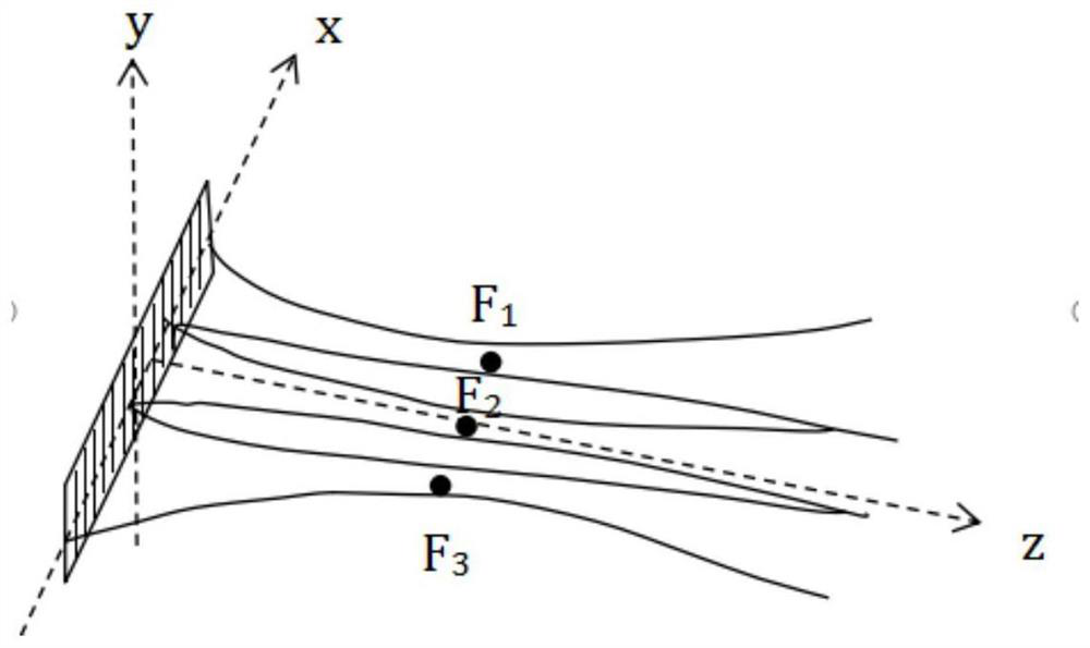
[0090] The focal sound pressure acquisition module 1 is used to obtain the sound pressure component p of the ultrasonic wave emitted by the ultrasonic probe at each focal position, wherein the required number of focal points, focal point positions and acoustic Pressure distribution, the focal point includes at least two; this module executes the method described in step S1 in Embodiment 1, which will not be repeated here.
[0091] The array element sound pressure acquisition module 2 is used to acquire the sound pressure h generated by each array element in the ultrasonic probe at each focus position; this module executes the method described in step S2 in Embodiment 1, and is not described here Let me repeat.
[0092] The sound pressure weight acquisition module 3 is configured to acquire the sound pressure weight w; the module executes the method d...
Embodiment 3
[0097] An embodiment of the present invention provides a computer device, such as Figure 9 As shown, the device may include a processor 51 and a memory 52, wherein the processor 51 and the memory 52 may be connected via a bus or in other ways, Figure 9 Take connection via bus as an example.
[0098]As a non-transitory computer-readable storage medium, the memory 52 can be used to store non-transitory software programs, non-transitory computer-executable programs and modules, such as corresponding program instructions / modules in the embodiments of the present invention. The processor 51 executes various functional applications and data processing of the processor by running the non-transitory software programs, instructions and modules stored in the memory 52, that is, implements the shear wave elastography method in the first method embodiment above.
[0099] The memory 52 may include a program storage area and a data storage area, wherein the program storage area may store...
PUM
 Abstract
Description
Claims
Application Information
 - R&D
- Intellectual Property
- Life Sciences
- Materials
- Tech Scout
- Unparalleled Data Quality
- Higher Quality Content
- 60% Fewer Hallucinations
Browse by: Latest US Patents, China's latest patents, Technical Efficacy Thesaurus, Application Domain, Technology Topic, Popular Technical Reports.
© 2025 PatSnap. All rights reserved.Legal|Privacy policy|Modern Slavery Act Transparency Statement|Sitemap|About US| Contact US: help@patsnap.com


