Liver blood vessel segmentation method based on CT image

A technology of liver blood vessels and CT images, applied in the field of medical image processing, can solve problems such as high signal-to-noise ratio, large differences between different individuals, and difficult to handle small blood vessel segmentation, and achieve the effect of improving the segmentation effect and improving the effect

Pending Publication Date: 2021-06-11
北京精诊医疗科技有限公司
View PDF4 Cites 4 Cited by
  • Summary
  • Abstract
  • Description
  • Claims
  • Application Information

AI Technical Summary

Problems solved by technology

Through CT, doctors can obtain a series of two-dimensional CT slices with enhanced blood vessels. However, abdominal CT images often have unfavorable factors such as low contrast, high signal-to-noise ratio, blurred boundaries, and adhesion between the liver and other tissues with similar gray levels. Due to the complex structure of liver vessels , blood vessels are entangled with each other, and there are great differences between different individuals. Liver blood vessel segmentation is facing great challenges
[0003] The existing 3D liver vessel segmentation methods can gene

Method used


Image

  • Liver blood vessel segmentation method based on CT image
  • Liver blood vessel segmentation method based on CT image
  • Liver blood vessel segmentation method based on CT image

Examples


Embodiment Construction

[0024] Below in conjunction with accompanying drawing and embodiment, technical solution of the present invention is described further:

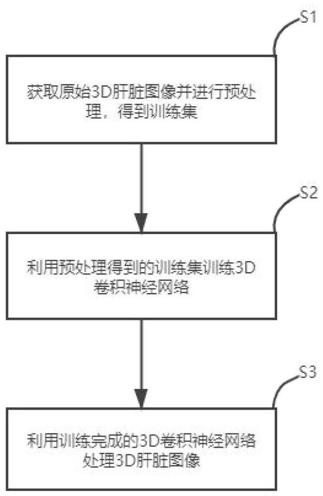
[0025] Provided in this embodiment is a liver vessel segmentation method based on CT images, such as figure 1 shown, including:

[0026] Step 1. Obtain the original 3D liver image and perform preprocessing to obtain the training set. In this embodiment, the original 3D liver image is adjusted for window width and level, and randomly cropped according to the patch size of 128×128×128, as a training set, in order to eliminate the uneven brightness distribution on the entire image problem, improving the contrast of blood vessels through illumination correction and smoothing CT image noise.

[0027] Step 2, using the obtained training set to train the convolutional neural network;

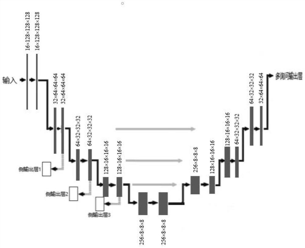
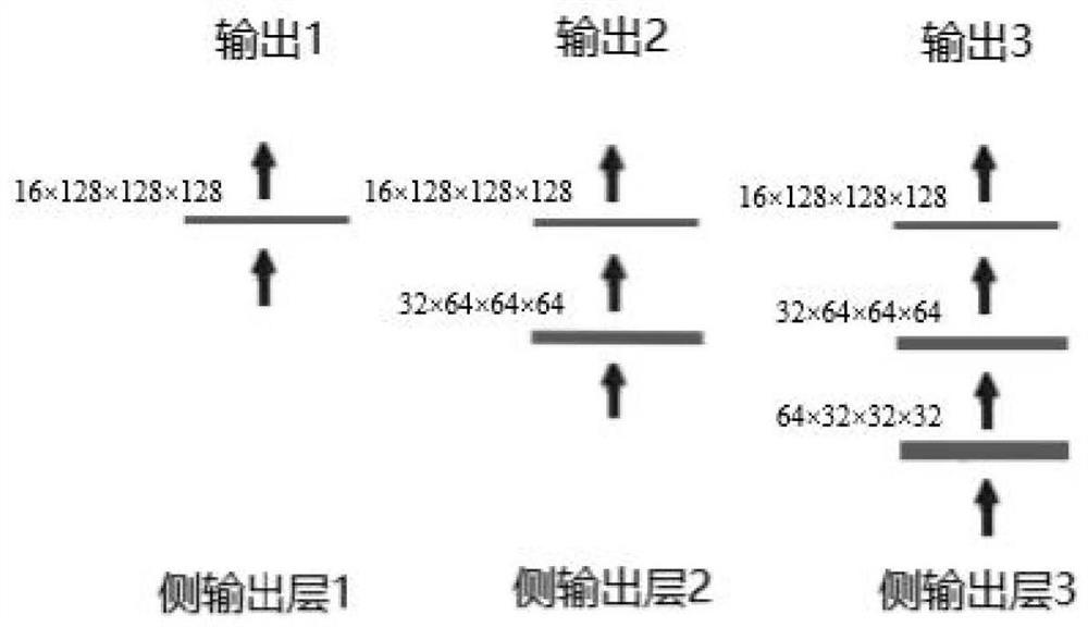
[0028] Such as figure 2 , the convolutional neural network in this embodiment adopts the Unet network structure, and the convolutional neural network has a tota...


PUM


Abstract

The invention discloses a liver blood vessel segmentation method based on a CT image, and the method comprises the steps of: firstly obtaining an original 3D liver image, and carrying out the preprocessing, and obtaining a training set; then using the training set obtained through preprocessing for training a 3D convolutional neural network, wherein the adopted 3D convolutional neural network adopts a Unet network structure, an encoder is provided with a side output layer for deeply supervising the structure of the convolutional network system, an output end is provided with two parallel branches, the upper branch is used for extracting features, different from the background, of the hepatic vein and the portal vein, and the lower branch is used for extracting features for distinguishing the hepatic vein and the portal vein; and finally processing the 3D liver image by using the trained 3D convolutional neural network to obtain a liver blood vessel segmentation result. According to the method, a side output layer is added from an encoder part to help bottom layer features to extract more semantic information, and meanwhile, two parallel branches are arranged at the output end, so that the segmentation effect of the hepatic vein and the portal vein in the liver image is improved.

Description

technical field [0001] The invention belongs to the field of medical image processing, and in particular relates to a liver blood vessel segmentation method based on CT images. Background technique [0002] Liver vascular segmentation and 3D reconstruction help to accurately obtain the overall information of abdominal liver vascular tissue, which is the premise of computer-aided liver disease diagnosis and liver surgery planning, and is of great significance for liver disease diagnosis and liver surgery guidance. CT (computed tomography) technology is one of the most commonly used diagnostic techniques for liver and blood vessel effects. Through CT, doctors can obtain a series of two-dimensional CT slices with enhanced blood vessels. However, abdominal CT images often have unfavorable factors such as low contrast, high signal-to-noise ratio, blurred boundaries, and adhesion between the liver and other tissues with similar gray levels. Due to the complex structure of liver ve...

Claims


Application Information

Patent Timeline
IPC IPC(8): G06T7/00G06T7/11G06T5/00G06N3/04G06N3/08
CPCG06T7/0012G06T7/11G06T5/002G06N3/08G06T2207/10081G06T2207/20081G06T2207/20084G06T2207/30056G06T2207/30101G06N3/045
Inventor 王博赵威申建虎张伟徐正清
Owner 北京精诊医疗科技有限公司
Why Eureka
Try Eureka