

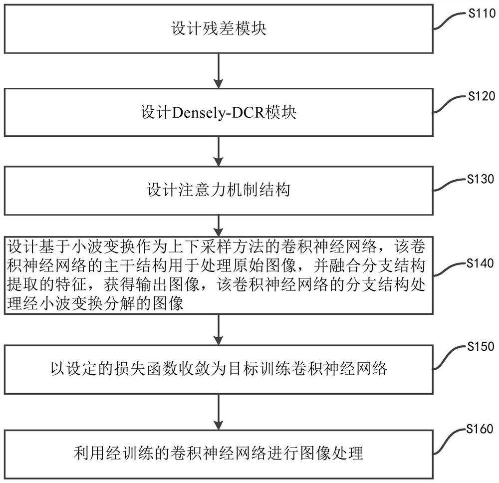
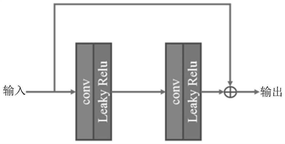
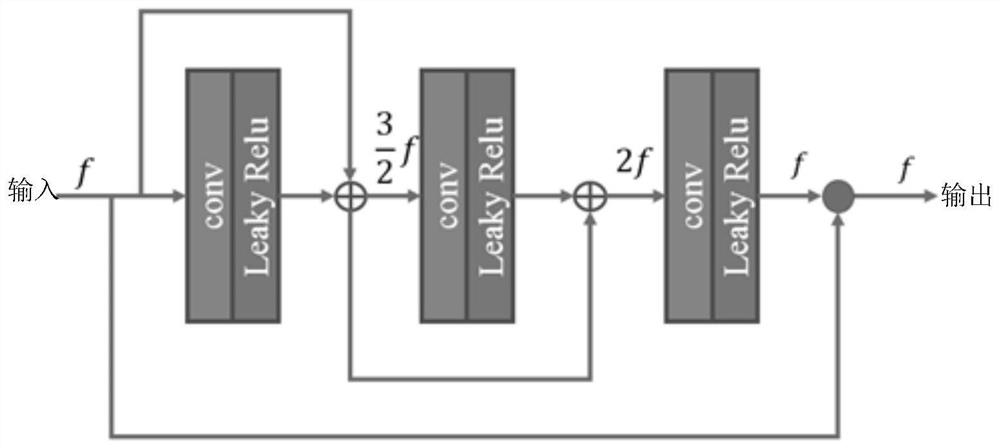
Deep learning method for medical image low-dose estimation
A medical image and deep learning technology, applied in the deep learning field of low dose estimation of medical images, can solve the problems of structural deformation, poor image reconstruction effect, large noise in PET images, etc., to improve the peak signal-to-noise ratio and strong generalization ability. , Enhance the effect of image detail information
- Summary
- Abstract
- Description
- Claims
- Application Information
AI Technical Summary
Problems solved by technology
Method used
Image

Examples
Embodiment Construction
[0024] Various exemplary embodiments of the present invention will now be described in detail with reference to the accompanying drawings. It should be noted that the relative arrangements of components and steps, numerical expressions and numerical values set forth in these embodiments do not limit the scope of the present invention unless specifically stated otherwise.
[0025] The following description of at least one exemplary embodiment is merely illustrative in nature and in no way taken as limiting the invention, its application or uses.
[0026] Techniques, methods and devices known to those of ordinary skill in the relevant art may not be discussed in detail, but where appropriate, such techniques, methods and devices should be considered part of the description.
[0027] In all examples shown and discussed herein, any specific values should be construed as exemplary only, and not as limitations. Therefore, other instances of the exemplary embodiment may have dif...
PUM
 Abstract
Description
Claims
Application Information