Method for establishing animal extrahepatic biliary tract malignant stenosis model
A method for establishing an animal liver, which is applied in the field of establishing a malignant stenosis model of the extrahepatic biliary tract in animals, can solve the problems of low tumor formation rate and difficulty in producing obstructive jaundice in animal models.
- Summary
- Abstract
- Description
- Claims
- Application Information
AI Technical Summary
Problems solved by technology
Method used
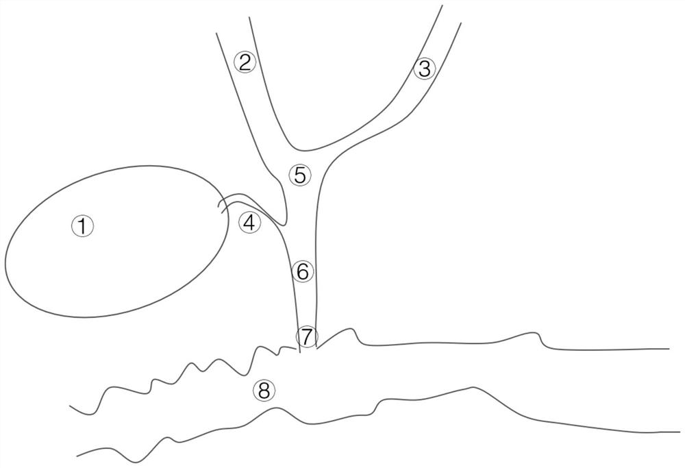
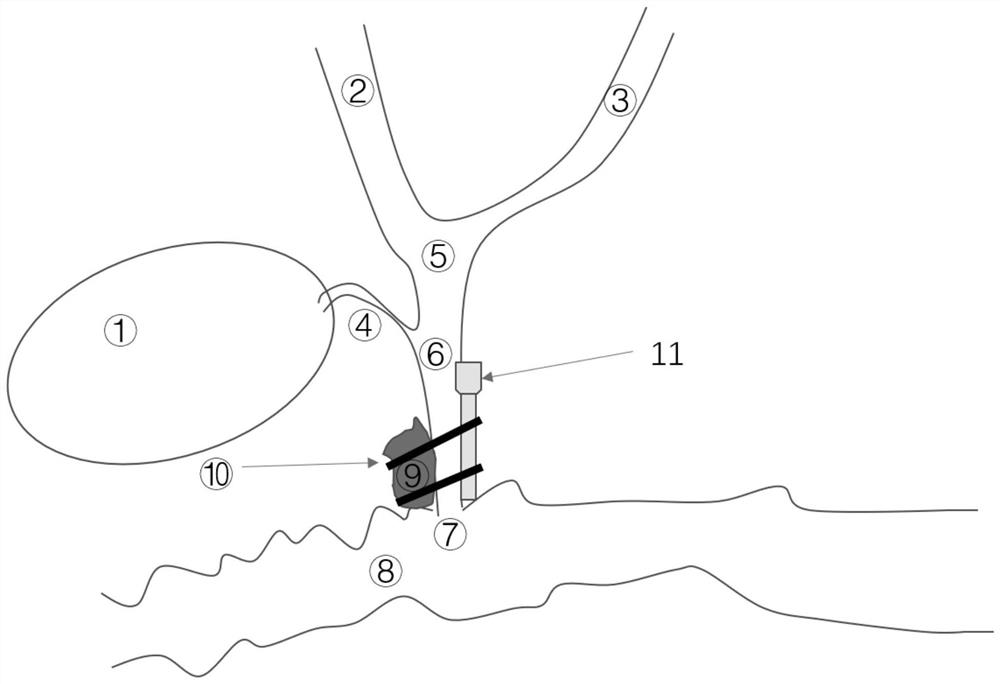
Image

Examples
Embodiment 1
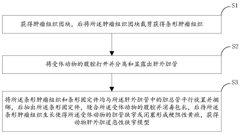
[0061] S1. Isolate from a donor rabbit with a tumor to obtain a tumor tissue mass, specifically:
[0062] The experimental animals were anesthetized through the ear vein, and the limbs of the experimental animals were fixed on the operating table in the supine position, the skin was prepared in the groin, and the towel was disinfected, and the rabbit tumor cells VX2 or tumor tissue were inoculated subcutaneously in the groin of the rabbit (the inoculation amount was 10 5 Suspension of tumor cells or more, 1mm 3 Tissue mass), about 2-3 weeks after inoculation, the tumor tissue in the groin area can grow to a diameter of about 1.5-20cm. Tumors were isolated and dissected under sterile conditions. The vigorously growing fish-like tissue at the edge of the tumor was cut out, placed in a plate filled with normal saline, the necrotic part, fiber, fat and other connective tissues were removed, and the remaining tumor tissue was trimmed into a strip-shaped tumor tissue with a size of...
Embodiment 2
[0066] S1. Isolate from a donor rabbit with a tumor to obtain a tumor tissue mass, specifically:
[0067] The experimental animals were anesthetized through the marginal ear vein, the limbs of the experimental animals were fixed on the operating table in the supine position, the skin was prepared in the groin area, and the towel was sterilized, and the rabbit tumor cells VX2 tumor cells or tumor tissues were inoculated subcutaneously in the groin area of the rabbits (the inoculation amount was 10 5 Suspension of tumor cells or more, 1mm 3 The above tissue mass), about 2-3 weeks after inoculation, the tumor tissue in the groin area can grow to a diameter of about 2cm. Tumors were isolated and dissected under sterile conditions. Cut out the vigorously growing fish-like tissue at the edge of the tumor, place it in a plate filled with normal saline, remove the necrotic part and connective tissue such as fiber and fat, and trim the remaining tumor tissue into a strip-shaped tum...
Embodiment 3
[0071] S1. Isolate from a donor rabbit with a tumor to obtain a tumor tissue mass, specifically:
[0072] The experimental animals were anesthetized through the marginal ear vein, the limbs of the experimental animals were fixed on the operating table in the supine position, the skin was prepared in the groin area, and the towel was sterilized, and the rabbit tumor cells VX2 tumor cells or tumor tissues were inoculated subcutaneously in the groin area of the rabbits (the inoculation amount was 10 5 Suspension of tumor cells or more or 1mm 3 The above tissue mass), about 2-3 weeks after inoculation, the tumor tissue in the groin area can grow to a diameter of about 2cm. Tumors were isolated and dissected under sterile conditions. Cut out the vigorously growing fish-like tissue at the edge of the tumor, place it in a plate filled with normal saline, remove the necrotic part and connective tissue such as fiber and fat, and trim the remaining tumor tissue into a strip-shaped t...
PUM
| Property | Measurement | Unit |
|---|---|---|
| Width | aaaaa | aaaaa |
| Length | aaaaa | aaaaa |
Abstract
Description
Claims
Application Information
 - R&D
- Intellectual Property
- Life Sciences
- Materials
- Tech Scout
- Unparalleled Data Quality
- Higher Quality Content
- 60% Fewer Hallucinations
Browse by: Latest US Patents, China's latest patents, Technical Efficacy Thesaurus, Application Domain, Technology Topic, Popular Technical Reports.
© 2025 PatSnap. All rights reserved.Legal|Privacy policy|Modern Slavery Act Transparency Statement|Sitemap|About US| Contact US: help@patsnap.com