Brain MRI image segmentation method and system

An MRI image and brain technology, applied in the field of medical image processing, can solve the problems of noise sensitivity and neglect of spatial information, and achieve the effect of effective and reliable segmentation, strong generalization ability, and strong anti-noise ability.

Active Publication Date: 2022-07-19
SHANXI UNIV
View PDF13 Cites 0 Cited by
  • Summary
  • Abstract
  • Description
  • Claims
  • Application Information

AI Technical Summary

Problems solved by technology

For MRI images of brain tissue, because different brain tissues such as white matter, gray matter, and cerebrospinal fluid are mixed together, the ambiguity of the transition area between tissues is more prominent, and the traditional hard-divided clustering method is no longer applicable.
In addition, previous clustering-based image segmentation methods mainly focus on the gray level information of a single pixel, ignoring the inherent spatial information in MRI images, and are more sensitive to noise.

Method used


Image

  • Brain MRI image segmentation method and system
  • Brain MRI image segmentation method and system
  • Brain MRI image segmentation method and system

Examples


Embodiment Construction

[0094] The technical solutions in the embodiments of the present invention will be described in detail below with reference to the embodiments of the present invention and the accompanying drawings. It should be pointed out that for those of ordinary skill in the art, without departing from the principle of the present invention, several modifications and improvements can also be made, which should also be regarded as belonging to the protection scope of the present invention.

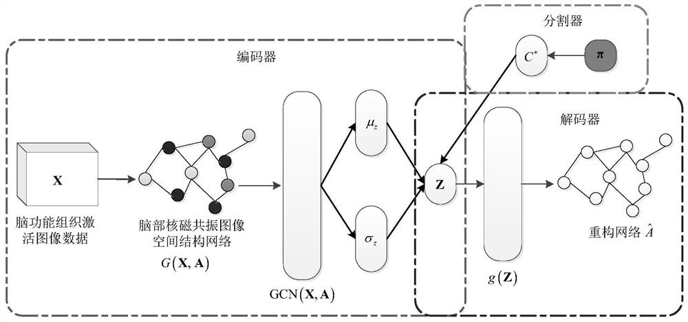
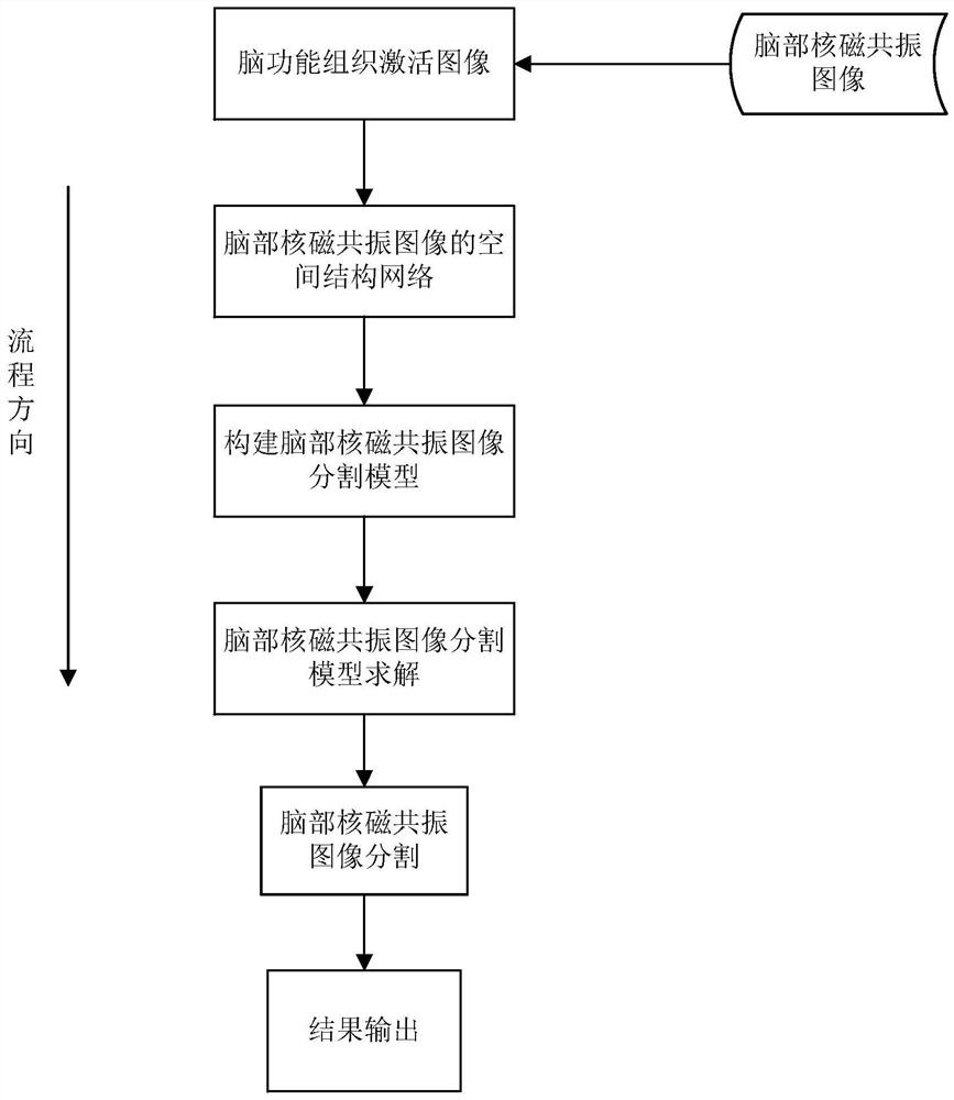
[0095] A brain MRI image segmentation method of the present invention is implemented by a computer program, and the following will be performed according to: image 3 The shown process details the specific implementation of the technical solution proposed by the present invention. Through the technical solution of the present invention, brain MRI images of subjects aged between 60 and 70 are selected from the MR Image Data dataset from Alzheimer's Disease Neuroimaging Initiative (ADNI) for image segmen...


PUM


Abstract

The invention relates to the technical field of medical image processing, and provides a brain nuclear magnetic resonance image segmentation method and system. The method mainly includes brain MRI image data preprocessing, brain MRI image segmentation model construction and optimization, brain MRI image segmentation and result output; the system includes a computer processor and memory, a brain MRI image preprocessing unit, Brain MRI image segmentation model training unit, brain MRI image segmentation and result output unit. The invention firstly constructs the spatial structure network of the brain MRI image, which can not only record the pixel point information of the brain functional tissue activation image, but also effectively express the spatial structure relationship between them, and then use the graph variational autoencoder structure to establish The brain MRI image segmentation model enables the model to have certain generation ability, and the model can obtain more robust and interpretable brain MRI image segmentation results.

Description

technical field [0001] The present invention relates to the technical field of medical image processing, and in particular, to a method and system for segmentation of a brain nuclear magnetic resonance image. Background technique [0002] Nuclear Magnetic Resonance Imaging (NMRI), also known as spin imaging, is to use the nuclear magnetic resonance phenomenon of a certain nucleus in human tissue to process the radio frequency signal obtained by electronic computer to reconstruct a certain level of the human body. Image diagnostic techniques. Magnetic resonance imaging is another major advancement in medical imaging after CT. Since its application in the 1980s, it has been rapidly developed and promoted. The main advantages of MRI technology include: a. Compared with other medical imaging techniques, the imaging contrast of soft tissue is higher; b. It is easier to qualitatively analyze a certain part of the human body and effectively find abnormal results; c. The human bod...

Claims


Application Information

Patent Timeline
Patent Type & Authority Patents(China)
IPC IPC(8): G06T7/00G06V10/26G06V10/74G06V10/82G06K9/62G06N3/04
CPCG06T7/0012G06T2207/10088G06T2207/30016G06V10/267G06N3/045G06F18/22
Inventor 杜航原王文剑白亮
Owner SHANXI UNIV
Why Eureka
Try Eureka