Blood vessel wall image segmentation method and device, computer equipment and storage medium

An image segmentation and blood vessel wall technology, applied in the field of medical imaging, can solve the problems of cumbersome registration operation process and easy introduction of errors.

Pending Publication Date: 2021-08-06
TSINGHUA UNIV
View PDF0 Cites 3 Cited by
  • Summary
  • Abstract
  • Description
  • Claims
  • Application Information

AI Technical Summary

Problems solved by technology

[0003] At present, the intracranial wall image segmentation scheme usually needs to manually draw the vessel centerline, or register with the angiographic image to obtain the vessel centerline mappin

Method used


Image

  • Blood vessel wall image segmentation method and device, computer equipment and storage medium
  • Blood vessel wall image segmentation method and device, computer equipment and storage medium
  • Blood vessel wall image segmentation method and device, computer equipment and storage medium

Examples


Embodiment Construction

[0062]In order to make the purpose, technical solution and advantages of the present application clearer, the present application will be further described in detail below in conjunction with the accompanying drawings and embodiments. It should be understood that the specific embodiments described here are only used to explain the present application, and are not intended to limit the present application.

[0063] The segmentation of vascular lumen based on MRI (Magnetic Resonance Imaging, nuclear magnetic resonance imaging) is a very challenging task. Noise and artifacts in MR (Magnetic Resonance, nuclear magnetic resonance) images are likely to cause low contrast between the vessel wall and surrounding tissues, complex The out-of-shape structure of the vascular lumen is also a technical difficulty to be considered in the segmentation. At present, traditional vascular wall image segmentation algorithms, such as region growing algorithm, fuzzy connection algorithm and multi-sc...


PUM


Abstract

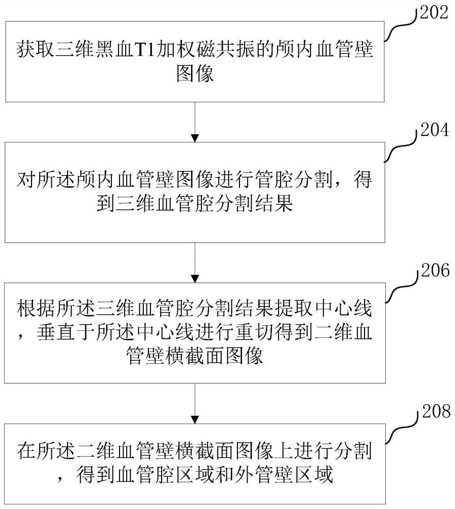
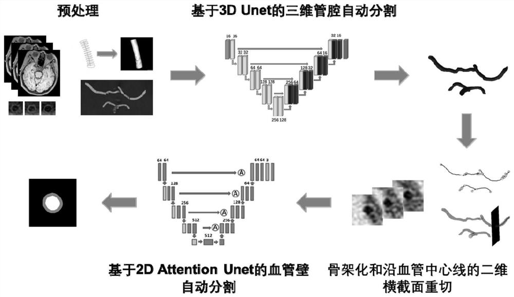
The invention relates to a blood vessel wall image segmentation method and device, computer equipment and a storage medium. The method comprises the following steps: acquiring an intracranial blood vessel wall image of three-dimensional black blood T1 weighted magnetic resonance; performing lumen segmentation on the intracranial vascular wall image to obtain a three-dimensional vascular lumen segmentation result; extracting a center line according to the three-dimensional blood vessel cavity segmentation result, and carrying out re-cutting perpendicular to the center line to obtain a two-dimensional blood vessel wall cross section image; and segmenting the two-dimensional blood vessel wall cross section image to obtain a blood vessel cavity area and an outer tube wall area. The three-dimensional lumen in the magnetic resonance image is firstly segmented, then the two-dimensional lumen is segmented on the two-dimensional blood vessel wall cross section image according to the three-dimensional lumen segmentation result, and the accuracy and the segmentation efficiency of blood vessel wall image segmentation are improved through accurate segmentation of the lumen and the lumen.

Description

technical field [0001] The present application relates to the technical field of medical imaging, in particular to a method, device, computer equipment and storage medium for segmenting a blood vessel wall image. Background technique [0002] Intracranial atherosclerosis is one of the most common causes of stroke. Vascular lumen stenosis determined by traditional angiographic imaging techniques is still the main method for diagnosing intracranial atherosclerosis, but intracranial atherosclerosis cannot be accurately judged only based on lumen stenosis. According to the study, the degree of stenosis of the vessel wall and the thickness of the vessel wall / area of ​​the vessel lumen are significantly related to plaque symptoms, so it is of great significance to accurately segment the intracranial vessel wall. [0003] At present, the intracranial wall image segmentation scheme usually needs to manually outline the vessel centerline, or register with the angiographic image to o...

Claims


Application Information

Patent Timeline
IPC IPC(8): G06T7/11G06T7/155G06T7/187
CPCG06T2207/10088G06T2207/20081G06T2207/30101G06T7/11G06T7/155G06T7/187
Inventor 陈慧军窦佳琦李雨泽赵锡海王雅洁
Owner TSINGHUA UNIV
Why Eureka
Try Eureka