Preparation method of collagen-based glucose fluorescent sensitive membrane
A collagen and glucose technology, applied in the field of fluorescent biosensors and preparation, can solve the problems of unpredictable hyperglycemia and hypoglycemia, reducing patient monitoring initiative, unfavorable long-term monitoring of blood sugar, etc., and achieves fewer preparation processes and safe and reliable preparation process. , the effect of low biological toxicity
- Summary
- Abstract
- Description
- Claims
- Application Information
AI Technical Summary
Problems solved by technology
Method used
Image

Examples
Embodiment 1
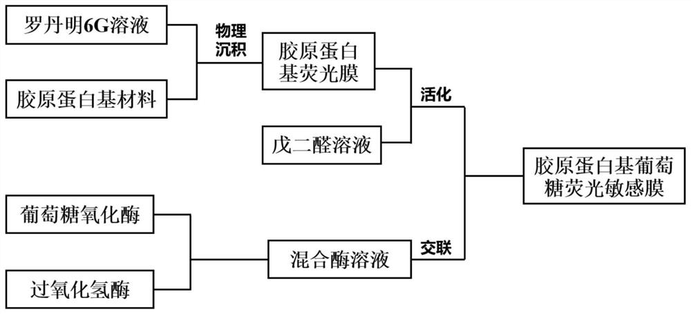
[0028] Prepare mixed enzyme solutions of glucose oxidase and catalase with enzyme activities of 800U / mL and 1600U / mL respectively, and store in a refrigerator at 4°C;
[0029] Cut the collagen base into small pieces of 10mm*10mm and immerse it in the rhodamine 6G solution with a concentration of 0.05mg / mL, take it out after soaking for 80 minutes, and rinse the remaining rhodamine 6G solution on the surface of the fluorescent film with deionized water. Dry at room temperature for later use;
[0030] Immerse the fluorescent film in a glutaraldehyde solution with a mass percentage of 1%, and take it out after soaking for 12 hours;
[0031] Place the activated fluorescent membrane on the surface of a clean glass slide, use a pipette to draw 150 μL of mixed enzyme solution, drop it on the surface of the fluorescent membrane, and add 150 μL of mixed enzyme solution dropwise after 5 hours of crosslinking, and the entire crosslinking The process is repeated 10 times.
Embodiment 2
[0033] Prepare mixed enzyme solutions of glucose oxidase and catalase with enzyme activities of 1000U / mL and 2000U / mL respectively, and store in a refrigerator at 4°C;
[0034] Cut the collagen base into small pieces of 12mm*12mm and immerse it in the rhodamine 6G solution with a concentration of 0.08mg / mL, take it out after soaking for 70 minutes, and rinse the remaining rhodamine 6G solution on the surface of the fluorescent film with deionized water. Dry at room temperature for later use;
[0035] Immerse the fluorescent film in a 0.5% glutaraldehyde solution by mass, and take it out after soaking for 21 hours;
[0036] Place the activated fluorescent membrane on the surface of a clean glass slide, use a pipette to draw 120 μL of mixed enzyme solution, drop it on the surface of the fluorescent membrane, add 120 μL of mixed enzyme solution dropwise after 6 hours of crosslinking, and the entire crosslinking The process was repeated 8 times.
Embodiment 3
[0038] Prepare mixed enzyme solutions of glucose oxidase and catalase with enzyme activities of 1200U / mL and 2400U / mL respectively, and store in a refrigerator at 4°C;
[0039] Cut the collagen base into small pieces of 13mm*13mm and immerse it in the rhodamine 6G solution with a concentration of 0.09mg / mL, take it out after soaking for 60 minutes, and rinse the remaining rhodamine 6G solution on the surface of the fluorescent film with deionized water. Dry at room temperature for later use;
[0040] Immerse the fluorescent film in a glutaraldehyde solution with a mass percentage of 0.2%, and take it out after soaking for 24 hours;
[0041] Place the activated fluorescent membrane on the surface of a clean glass slide, use a pipette to draw 100 μL of mixed enzyme solution, drop it on the surface of the fluorescent membrane, add 100 μL of mixed enzyme solution dropwise after 8 hours of crosslinking, and the entire crosslinking The process was repeated 6 times.
PUM
 Abstract
Description
Claims
Application Information
 - R&D Engineer
- R&D Manager
- IP Professional
- Industry Leading Data Capabilities
- Powerful AI technology
- Patent DNA Extraction
Browse by: Latest US Patents, China's latest patents, Technical Efficacy Thesaurus, Application Domain, Technology Topic.
© 2024 PatSnap. All rights reserved.Legal|Privacy policy|Modern Slavery Act Transparency Statement|Sitemap