PET scanning system and scanning method

A scanning method and scanning system technology, which can be used in computer tomography scanners, medical science, and instruments for radiological diagnosis, etc., can solve the problems of inability to realize metabolic monitoring, PET imaging cannot reflect the metabolic status, adjustment and coordination difficulties, etc. Achieve the effect of improving image reconstruction quality, solving structural coordination difficulties, and ensuring temporal resolution

Pending Publication Date: 2021-08-20
SHANGHAI JIAO TONG UNIV
View PDF0 Cites 0 Cited by
  • Summary
  • Abstract
  • Description
  • Claims
  • Application Information

AI Technical Summary

Problems solved by technology

There are many defects in the existing system: first, PET scanning needs the assistance of gas intubation anesthesia, respiratory gating, heat preservation and other processes, and it is difficult to adjust and cooperate; second, during the scanning process, the animal is under anesthesia, and PET imaging cannot reflect the natural state The metabolism in the lower body; the third is that the system cannot realize the metabolic monitoring of animals in the state of exercise, and the application has limitations

Method used


Image

  • PET scanning system and scanning method
  • PET scanning system and scanning method
  • PET scanning system and scanning method

Examples


Embodiment 1

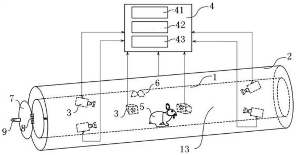
[0045] figure 1 It is a linear PET scanning system with circular tubular structure, which can be used for animal scanning. The PET scanning system includes a limiting device 1 for limiting the range of motion of a scanning object 5 , a PET detector array 2 , a positioning device 3 and a signal processing device 4 .

[0046] The restricting device 1 is in the shape of a straight circular tube, which is provided with a lumen 13 . The lumen 13 is linear and provides space for the scanning object 5 to move. One end of the restricting device 1 is closed, and the other end has a first door 7 that can be opened. The first door 7 is circular. The first door 7 is connected to the restricting device 1 through a hinge 8 , and is opened and closed through a hasp 9 . There is an opening 6 on the upper side of the restriction device 1, and the opening 6 communicates with the lumen 13, which is used as a vent or a window for applying stimulation. Open the first door 7 to place the scann...

Embodiment 2

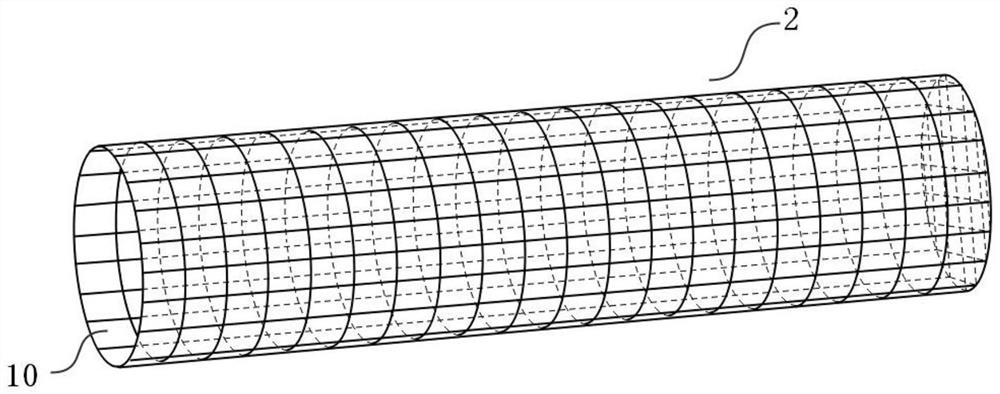
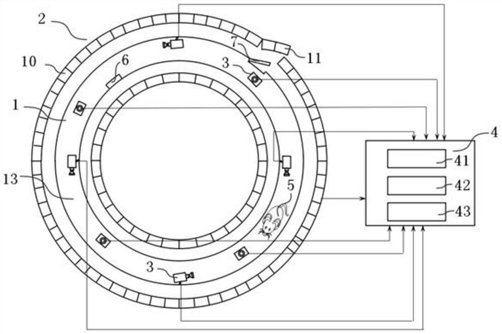
[0050] image 3 It is a top sectional view of a ring-shaped PET scanning system. The PET scanning system includes a PET detector 10 . A plurality of PET detectors 10 are formed as Figure 4 The three-dimensional annular PET detector array 2 is shown. A second door 11 is arranged on the PET detector array 2 , and the second door 11 is arranged so that it can be opened and closed. Open the second door 11 to enter the inside of the PET detector array 2 . Closing the second door 11 can seal the PET detector array 2 . The PET detector array 2 is surrounded by a ring-shaped restricting device 1 . The restriction device 1 has a circular lumen 13 . The lumen 13 is the activity space of the scanning object 5 . The restriction device 1 is provided with a first door 7 . The first door 7 is provided so as to be openable and closable. Open the first door 7 to enter the lumen 13 . Closing the first door 7 can seal the lumen 13 . The restriction device 1 is also provided with an o...

Embodiment 3

[0055] Figure 5 is a side view cross-sectional view of an irregular structure PET system for animals. The PET detector 10 is formed as Figure 6 Shown is a PET detector array 2 of irregular structure with one end open and the other end closed, which surrounds the limiting device 1 . The shape of the confinement device 1 is the same as that of the PET detector array 2 . The restriction device 1 consists of a horizontal section and a slope section. Before scanning starts, the first door 7 on the restriction device 1 is opened, the scanning object 5 injected with radiopharmaceuticals is put into the restriction device 1 , and then the first door 7 is closed. Eleven lasers 12 are fixed on the inner surface of the restricting device 1 , and are respectively located on the top and side of the straight section of the restricting device 1 . The orientation of the laser 12 on the top of the restriction device 1 is approximately parallel to the axis of the restriction device 1 , an...


PUM


Abstract

The invention relates to a PET (Positron Emission Tomography) scanning method. The PEC scanning method comprises the following steps of: Step 31, injecting radiopharmaceutical into a scanned object, and placing the scanned object in a limiting device for limiting a motion range; Step 32, acquiring a signal containing animal position and attitude information by adopting positioning equipment and outputting the signal; Step 33, processing the signal in the step 32 by using a signal processing device so as to obtain three-dimensional position and attitude of a world coordinate system where the scanned object is located; Step 34,detecting the radiation condition in the scanned object by a PET detector and outputting the signal according to the detection result; and step 35, in the scanning process, generating multi-frame PET data and three-dimensional coordinates and attitude data of the scanned object on a time axis, and processing the data to generate a PET image. The invention further relates to a PET scanning system which comprises the limiting device, the PET detector, positioning equipment and a computer. The defects that in the prior art, all structures are difficult to cooperate, PET imaging is only limited to an anesthesia state, and the application range is narrow, are overcome.

Description

technical field [0001] The invention relates to a PET scanning system and a scanning method. Background technique [0002] At present, animal in vivo imaging is widely used in the research of tumor formation mechanism, functional metabolism, new drug development and other fields. The establishment of animal models has important reference significance for the study of human disease mechanism. With the development of imaging technology, Positron Emission Tomography (PET, Positron Emission Tomography) is playing an increasingly important role in functional imaging. [0003] The process of PET technology is as follows: first inject radiopharmaceuticals (for example, fluorodeoxyglucose FDG) into the living body, and the drug gathers in the metabolically active area in the body and reflects it through the image. Specifically, the decay of radionuclide releases positrons, which collide with nearby negative electrons to annihilate, producing a pair of photons with energy 511keV in ...

Claims


Application Information

Patent Timeline
Patent Type & Authority Applications(China)
IPC IPC(8): A61B6/03A61B6/00
CPCA61B6/037A61B6/508A61B6/5264
Inventor 赵俊周玉府
Owner SHANGHAI JIAO TONG UNIV
Why Eureka
Try Eureka