Multi-modal retina image fusion method and system based on image registration

An image registration and image fusion technology, applied in the field of image processing, can solve the problems of no constraints, inaccurate image space transformation, inaccurate image control transformation, etc., to assist diagnosis and treatment, and ensure stability and accuracy. Effect

Pending Publication Date: 2021-08-24
GUANGDONG GENERAL HOSPITAL
View PDF5 Cites 5 Cited by
  • Summary
  • Abstract
  • Description
  • Claims
  • Application Information

AI Technical Summary

Problems solved by technology

First, due to the specificity of retinal images (repetitive structure, multimodality), inaccurate feature extraction and description can generate a large number of false feature matches
In addition, in the process of image space transformation, the above research does not use constraints or only uses a single constraint method, which will lead to inaccurate image space transformation
These issues will affect the robustness and accuracy of multimodal retinal image fusion based on image registration
[0006] In summary, the existing retinal image registration methods generally suffer from inaccurate feature extraction and description, which lead to a large number of erroneous feature matches, and the constraint method leads to inaccurate transformation of image controls, which in turn affects the robustness and accuracy of multimodal retinal image fusion. precision

Method used


Image

  • Multi-modal retina image fusion method and system based on image registration
  • Multi-modal retina image fusion method and system based on image registration
  • Multi-modal retina image fusion method and system based on image registration

Examples


Embodiment 1

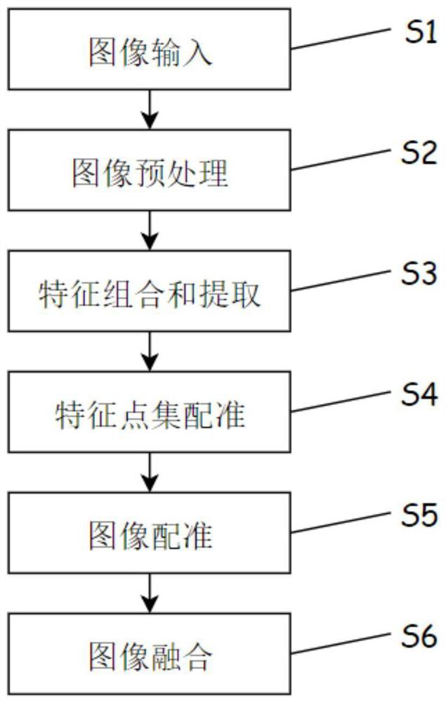
[0124] This embodiment proposes a multi-modal retinal image fusion method based on image registration, as shown in the attached Figure 1-6 shown. The process steps are as attached in the manual figure 1 , the specific scheme is as follows:

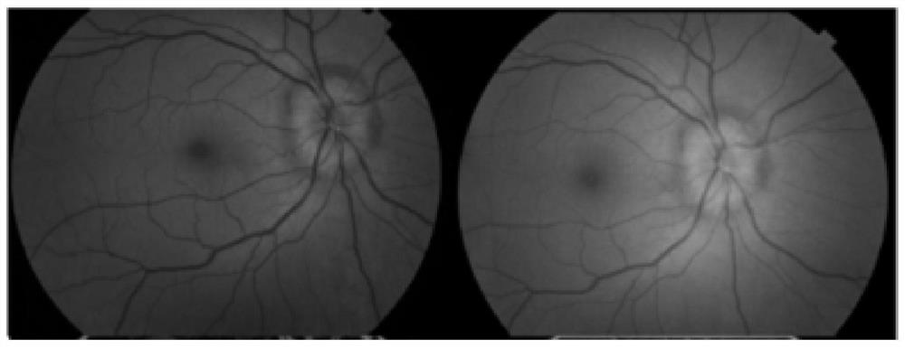
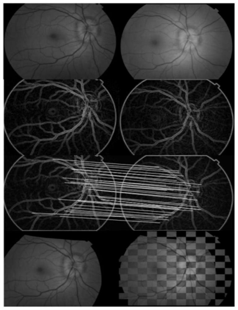
[0125] S1. Image input: acquiring a retinal image pair including a source image and a target image;

[0126] S2. Image preprocessing: Preprocessing the retinal image pair, obtaining the retinal edge image pair, extracting a feature point set from each edge image pair, the feature point set includes the source feature point set extracted from the preprocessed source image and the set of target feature points extracted from the preprocessed target image;

[0127] S3. Feature combination and extraction: combine multiple feature descriptors to construct a multi-feature difference descriptor, and guide the feature extraction of feature point sets;

[0128] S4. Feature point set registration: Evaluate the correspondence between the source f...

Embodiment 2

[0233] In this embodiment, on the basis of Embodiment 1, a multimodal retinal image fusion method based on image registration proposed in Embodiment 1 is modularized to form a multimodal retinal image fusion system based on image registration. The schematic diagram of each module is attached to the manual Figure 7 shown.

[0234] A multimodal retinal image fusion system based on image registration, comprising sequentially connected image input unit 1, image preprocessing unit 2, feature combination and extraction unit 3, feature point set registration unit 4, image registration unit 5 and Image fusion unit6.

[0235] Image input unit 1: for acquiring a retinal image pair including a source image and a target image. Each pair of retinal images includes a source image and a target image.

[0236] Image preprocessing unit 2: used to preprocess retinal image pairs, obtain retinal edge image pairs, and extract feature point sets from each pair of retinal edge image pairs. The f...


PUM


Abstract

The invention provides a multi-modal retina image fusion method and system based on image registration. The method comprises the steps of obtaining a retina image pair; preprocessing, obtaining retina edge image pairs, and extracting a feature point set from each retina edge image pair; combining a plurality of feature descriptors to construct a multi-feature difference descriptor, and guiding feature extraction of the feature point set; updating spatial transformation of the source feature point set according to the corresponding relation matrix; performing image registration, and obtaining an image after spatial transformation in combination with the target image; and fusing the image after spatial transformation and a preset reference image to obtain a fused image. According to the method, the defects of insufficient image registration precision and inaccurate image space transformation in a multi-modal retina image fusion process are overcome, and high-precision and high-stability registration performance can be realized. On the basis of high-precision image registration, effective multi-modal eye fundus image fusion is carried out, so that an ophthalmologist can conveniently observe the change of a lesion area.

Description

technical field [0001] The present invention relates to the field of image processing, in particular, to a multimodal retinal image fusion method and system based on image registration. Background technique [0002] Fundus retinal images are an important basis for the diagnosis of various retinal diseases including glaucoma and age-related macular degeneration. In addition, the fundus, as the only direct-viewing vascular window in the human body, can reflect the hemodynamic changes of other organs in the whole body to a certain extent. Multimodal retinal images usually contain important retinal local structure and temporal information. If the retinal images of different modalities of the same patient can be fused, it can provide doctors with a variety of complementary information on the lesion, thereby providing a more comprehensive and clear basis for doctors to make decisions about diagnosis and treatment. [0003] Due to the complex structure of the vascular network in ...

Claims


Application Information

Patent Timeline
Patent Type & Authority Applications(China)
IPC IPC(8): G06T5/50G06T7/00G06T7/33G06K9/46
CPCG06T5/50G06T7/33G06T7/0012G06T2207/20221G06T2207/30041G06V10/462
Inventor 余洪华蔡宏民但婷婷刘宝怡肖宇方莹
Owner GUANGDONG GENERAL HOSPITAL
Why Eureka
Try Eureka