
Method and system for identifying renal tubular atrophy area based on deep learning

A technology of area recognition and deep learning, which is applied in image data processing, instruments, calculations, etc., can solve the problems of medical needs and resource medical means that cannot meet patients, consume energy, etc., achieve intuitive display results, improve work efficiency, and detect high precision effect

Active Publication Date: 2021-08-27
TAIYUAN UNIV OF TECH
View PDF6 Cites 0 Cited by
  • Summary
  • Abstract
  • Description
  • Claims
  • Application Information

AI Technical Summary

Problems solved by technology

Renal tubular atrophy is the main form of chronic kidney disease. When checking whether a patient suffers from renal tubular atrophy, a pathologist is required to observe the patient's kidney pathology section, and an authoritative pathologist is required to judge the area ratio of the atrophic area. However, medical needs and resources The contradiction between the traditional medical methods can not meet the needs of patients
The pathology departments of some tertiary hospitals produce thousands of kidney pathology sections every day. Although most of them may not have positive results, doctors still need to strictly examine each pathology section under a microscope to determine whether there is atrophy in the renal tubules, which consumes a lot of energy.

Method used


Image

  • Method and system for identifying renal tubular atrophy area based on deep learning
  • Method and system for identifying renal tubular atrophy area based on deep learning
  • Method and system for identifying renal tubular atrophy area based on deep learning

Examples


Embodiment 1

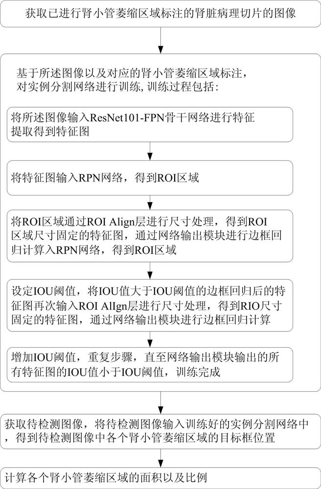
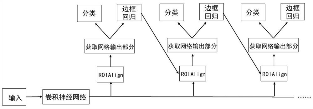
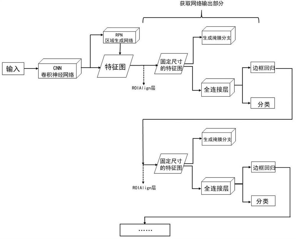
[0058] Such as Figure 1~3 As shown, Embodiment 1 of the present invention provides a method for identifying a renal tubular atrophy region based on deep learning, including the following steps:

[0059] S1. Obtain an image of a pathological section of the kidney that has been marked with a region of renal tubular atrophy.

[0060] Specifically, the operator first fixes the kidney pathological slides on the fully automatic digital pathological slide scanner, and then the scanner digitizes the pathological slides. Due to the large memory of the digitized pictures, the code reads the data too slowly when running , so each picture is cut into several small pictures of the same size in turn, and the cut pictures are named according to the rule of "patient number_serial number", such as Figure 4 shown. Then input the digitized pictures into the image clarity evaluation algorithm for screening. Use labelme labeling software to manually label the renal tubular atrophy area on the...

Embodiment 2

[0081] Such as Figure 7 As shown, Embodiment 2 of the present invention provides a system for identifying renal tubular atrophy regions based on deep learning, including: an acquisition unit, a training unit, a detection unit, and a calculation unit.

[0082] Wherein, the acquiring unit is used to acquire the image of the renal pathological section marked with the renal tubular atrophy region; the training unit is used to train the instance segmentation network based on the image and the corresponding renal tubular atrophy region marked. The detection unit is used to obtain the image to be detected, input the image to be detected into the trained instance segmentation network, and obtain the target frame position of each tubular atrophy area in the image to be detected; the calculation unit is used to calculate the area of ​​each tubular atrophy area and Proportion.

[0083] Specifically, the training process includes:

[0084] The image is input to the ResNet101-FPN backbo...


PUM


Abstract

The invention belongs to the technical field of artificial intelligence assisted medical treatment, and particularly relates to a method and a system for identifying a renal tubular atrophy area based on deep learning, and the method comprises the following steps: S1, obtaining an image of a kidney pathological section subjected to renal tubular atrophy area labeling; S2, training an instance segmentation network based on the image and a corresponding renal tubular atrophy area label, wherein the instance segmentation network is an improved mask-RCNN, a cascade network is added to the network for cascading each detection model, an IOU threshold value is set to define a sample training model, the output of the previous detection model is used as the input of one detection model, and the IOU value rises all the time; S3, inputting a to-be-detected image into the trained instance segmentation network to obtain a target frame position of each renal tubular atrophy area in the to-be-detected image; and S4, calculating the area and proportion of each renal tubular atrophy area. According to the invention, the detection of the renal tubular atrophy area can be realized, the detection precision is high, and the omission ratio is low.

Description

technical field [0001] The invention belongs to the technical field of artificial intelligence-assisted medical treatment, and in particular relates to a method and system for identifying renal tubular atrophy regions based on deep learning. Background technique [0002] Chronic kidney disease (CKD) is currently recognized by the World Health Organization (WHO) as one of the major public health challenges, and renal biopsy plays a key role in the diagnosis and treatment of CKD. At present, the pathological diagnosis of renal biopsy has become an important reference for the diagnosis, treatment and prognosis of patients with renal diseases. [0003] Pathology plays a vital role in the medical field. Among them, in the diagnosis of chronic kidney disease, the diagnostic results of the kidney tissue after coarse needle puncture, forceps, cutting, and resection are the most accurate and reliable after being made into pathological sections. "gold standard". Many kidney diseases...

Claims


Application Information

Patent Timeline
IPC IPC(8): G06T7/00G06T7/13G06T7/136G06T7/62G06T7/187
CPCG06T7/0012G06T7/13G06T7/136G06T7/62G06T7/187G06T2207/20104G06T2207/20081G06T2207/30084
Inventor 李明赖叶鑫王晨郝芳李心宇刘雪宇
Owner TAIYUAN UNIV OF TECH
Learn More