Acid-fast staining method and quality monitoring method
A technology for acid-resistant dyeing and quality monitoring, which is applied in the direction of measuring devices, instruments, and analytical materials, and can solve problems such as inability to decolorize
- Summary
- Abstract
- Description
- Claims
- Application Information
AI Technical Summary
Problems solved by technology
Method used
Image
Examples
Embodiment 1
[0037] Exploration of the causes of inability to decolorize in acid-fast staining.
[0038] 1. Find the problem.
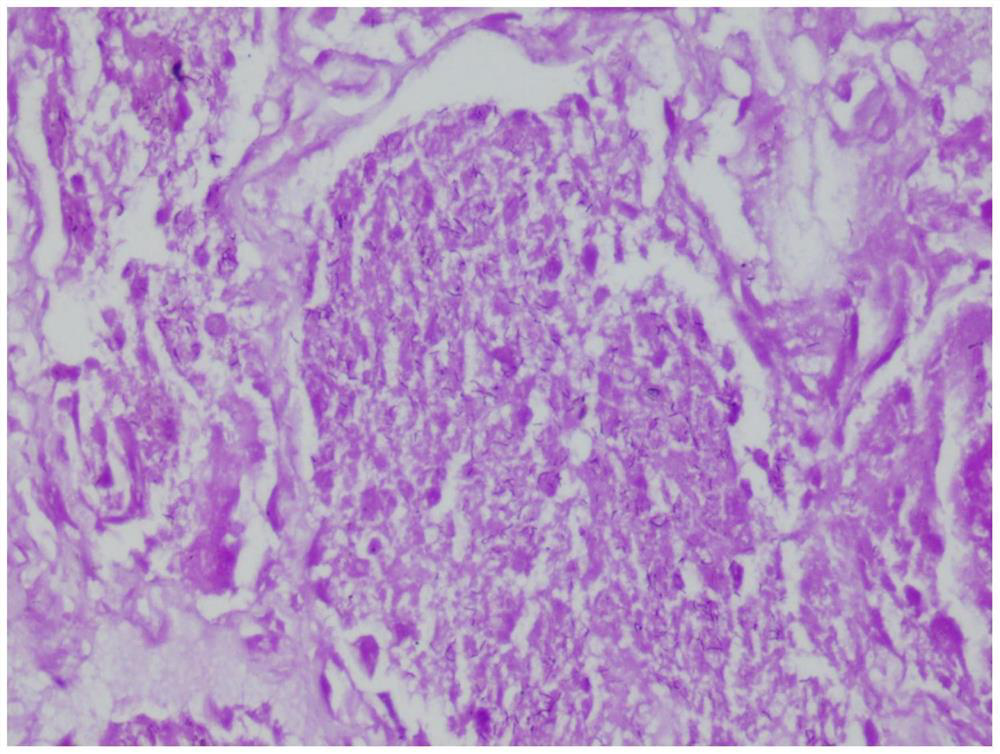
[0039] The inventor carried out acid-fast staining according to the conventional method, and found that the background of acid-fast staining sometimes appeared to be red-stained, which could not be removed by 20% sulfuric acid decolorization. figure 1 As shown, the tissue section is generally purple-red, the background is not well differentiated, and the contrast is poor, making it difficult to distinguish whether there are mycobacteria.
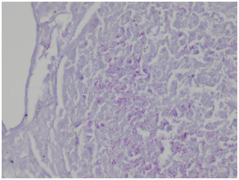
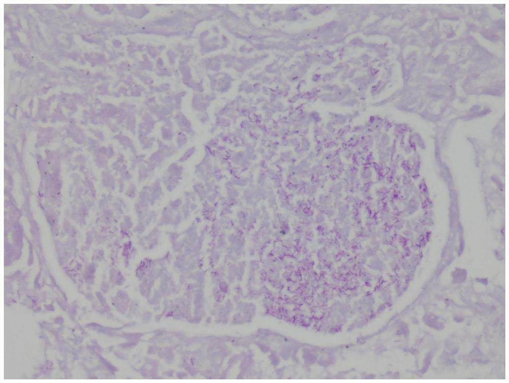
[0040] After re-slicing and staining, the section was slightly reddish under the microscope, as shown in the microscope. figure 2 As shown, Mycobacterium tuberculosis is red in number and easy to observe. The background is light blue and the contrast is clear.
[0041] 2. Preliminary screening of the reasons.
[0042] In order to find the root of the problem systematically, we list the key links and elements in the experimen...
Embodiment 2
[0060] Study on the standard of formaldehyde content in the quality control of acid-fast dyeing.
[0061] 1. Method.
[0062] During the experiment, the environmental formaldehyde concentration monitoring link was set up to compare the effects of different environmental formaldehyde concentrations on acid staining.
[0063] 2. Results.
[0064] After sorting out the experimental results, it was found that when the concentration of formaldehyde in the environment is less than 0.20mg / m 3 When the section is stained, it can be successfully decolorized and differentiated with an acid decolorizer. After the section is washed with water, the tissue is slightly light red, and the background is clean under the microscope, and Mycobacterium tuberculosis can be clearly observed; when the concentration of formaldehyde in the environment is greater than 0.20mg / m 3 The red background of the section cannot be decolorized with acid decolorizer. After the section is washed with water, the...
Embodiment 3
[0067] A method for monitoring the quality of acid-fast staining, including a formaldehyde detection step in the staining incubation process, specifically as follows:
[0068] 1. Method: Put the formaldehyde detection reagent in the corner of the incubation box (transparent, with a cover, convenient for observing the situation in the box), place the absorption box in the formaldehyde detection kit (brand: Lvzhiyuan, model: Z-0434), Then pour all the detection solution in the formaldehyde detection kit into the absorption box and mix well. Cover the lid of the incubation box and wait for the reagents to react for 30 minutes. Pour the chromogen in the formaldehyde detection kit into the absorption box, mix well, and let stand for 10 minutes.
[0069] 2. Observation results: Use the absorption box and the color comparison card in the formaldehyde detection kit to perform color comparison, and obtain the result of the formaldehyde concentration in the incubation box.
[0070] 3....
PUM
 Abstract
Description
Claims
Application Information
 - R&D
- Intellectual Property
- Life Sciences
- Materials
- Tech Scout
- Unparalleled Data Quality
- Higher Quality Content
- 60% Fewer Hallucinations
Browse by: Latest US Patents, China's latest patents, Technical Efficacy Thesaurus, Application Domain, Technology Topic, Popular Technical Reports.
© 2025 PatSnap. All rights reserved.Legal|Privacy policy|Modern Slavery Act Transparency Statement|Sitemap|About US| Contact US: help@patsnap.com


