Reticulocyte detection dye, detection reagent, preparation method of detection reagent, detection method of sample analyzer and sample analyzer
A technology of reticulocytes and detection reagents, which is applied in the field of blood cell analysis and can solve problems such as the inability to meet the requirements of automated testing
- Summary
- Abstract
- Description
- Claims
- Application Information
AI Technical Summary
Problems solved by technology
Method used
Image
Examples
preparation example Construction
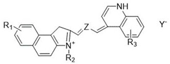
[0052] The present invention also provides a method for preparing a reticulocyte detection reagent, the method comprising: after configuring the above-mentioned detection dye and the diluent in a certain proportion to a preset volume, stirring evenly at room temperature, using 0.22 um filter membrane for filtration, and the filtrate after filtration is tested for the sample.
[0053] The present invention also provides a detection method for a sample analyzer. The method includes the following steps: using the reticulocyte detection reagent as described above, treating the sample with the detection reagent, and the detection reagent transforms the reticulocyte in the sample into a spherical shape. to enable the nucleic acid of the reticulocytes to specifically combine with the fluorescent dye, thereby realizing the counting of the reticulocytes.
[0054] The present invention also provides a sample analyzer adopting the detection method of the sample analyzer, the sample analy...
Embodiment example 1
[0058] Reagent name concentration Phosphate buffer pair 3.6g / L sodium hydroxide 2.1 g / L sodium sulfate 0.8g / L Decyl sultaine 1.1 g / L Octyltrimethylammonium Chloride 1.6g / L Isothiazolinone 0.04g / L pure water Dilute to 1000mL
[0059] Reagent name concentration Dye 1 0.0035 g Isopropanol 3.0 g Ethylene glycol Quantitative to 100g
[0060] Prepare the diluent and detection dye as shown in the above table and dilute to the specified amount, stir evenly at room temperature, and filter with a 0.22um filter membrane. Use the Sysmex XN1000 instrument to compare the supporting reagents with the self-made reagent models. See Table 1 for details of the results.
[0061] Table 1:
[0062] RET% Invention RET%XN-1000 Absolute deviation (no more than ±0.3) LFR Invention LFR XN-1000 Relative deviation (no more than ±30%) MFR Invention MFR XN-1000 Relative deviation (no...
Embodiment example 2
[0065] Reagent name concentration Phosphate buffer pair 3.6g / L sodium hydroxide 2.1 g / L sodium sulfate 0.8g / L Decyl sultaine 1.1 g / L Octyltrimethylammonium Chloride 1.6g / L Isothiazolinone 0.04g / L pure water Dilute to 1000mL
[0066] Reagent name concentration Dye 2 0.003 g Isopropanol 3.0 g Ethylene glycol Quantitative to 100g
[0067] Prepare the diluent and detection dye as shown in the above table and dilute to the specified amount, stir evenly at room temperature, and filter with a 0.22um filter membrane. Use the Sysmex XN1000 instrument to compare the supporting reagents with the self-made reagent models. See Table 2 for details of the results.
[0068] Table 2:
[0069] RET% Invention RET%XN-1000 Absolute deviation (no more than ±0.3) LFR Invention LFR XN-1000 Relative deviation (no more than ±30%) MFR Invention MFR XN-1000 Relative deviation (no ...
PUM
 Abstract
Description
Claims
Application Information
 - R&D
- Intellectual Property
- Life Sciences
- Materials
- Tech Scout
- Unparalleled Data Quality
- Higher Quality Content
- 60% Fewer Hallucinations
Browse by: Latest US Patents, China's latest patents, Technical Efficacy Thesaurus, Application Domain, Technology Topic, Popular Technical Reports.
© 2025 PatSnap. All rights reserved.Legal|Privacy policy|Modern Slavery Act Transparency Statement|Sitemap|About US| Contact US: help@patsnap.com


