SnS2 microsphere SERS substrate with capillary effect as well as preparation method and application of SnS2 microsphere SERS substrate

A microsphere, na2sno3 3h2o technology, applied in the field of laser Raman spectroscopy and detection, can solve the problems of low sensitivity and unfavorable probe molecule convergence phenomenon, etc.

Active Publication Date: 2021-10-08
SHANGHAI INST OF CERAMIC CHEM & TECH CHINESE ACAD OF SCI
View PDF8 Cites 0 Cited by
  • Summary
  • Abstract
  • Description
  • Claims
  • Application Information

AI Technical Summary

Problems solved by technology

However, the smooth surface of pure SnS2 nanosheets has no wrinkles, which is not conducive to the aggregation of probe molecules on the substrate surface at low concentrations, resulting in low sensitivity as a SERS substrate.

Method used


Image

  • SnS2 microsphere SERS substrate with capillary effect as well as preparation method and application of SnS2 microsphere SERS substrate
  • SnS2 microsphere SERS substrate with capillary effect as well as preparation method and application of SnS2 microsphere SERS substrate
  • SnS2 microsphere SERS substrate with capillary effect as well as preparation method and application of SnS2 microsphere SERS substrate

Examples


Embodiment 1

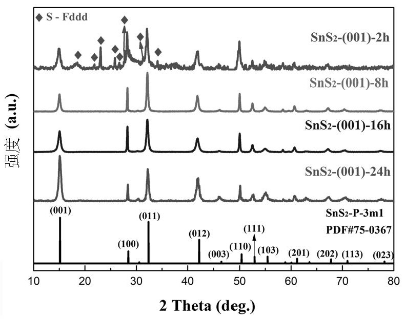
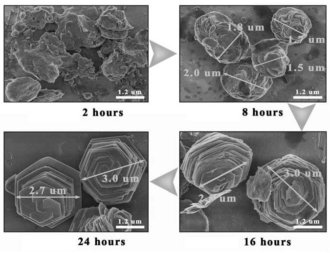
[0058] First, 1.6 g of TTA powder (21.3 mmoL) was added to 55 mL of deionized water, and placed under electromagnetic stirring at a temperature of 60° C. for about 10 minutes to rapidly dissolve the powder to obtain a mixed transparent solution. Then, add 0.8g Na 2 SnO 3 ·3H 2 O powder (3mmoL) was added to the above TTA solution, and electromagnetic stirring was continued at 60°C for about 20 minutes to dissolve the powder quickly and mix uniformly to obtain a precursor solution. Then the above precursor solution was placed in a 100mL PPL-lined hydrothermal reaction kettle, and hydrothermally reacted at 180°C for 24 hours to obtain a brown precipitate, as Figure 11 shown. Finally, the brown precipitate was centrifuged, washed three times with deionized water, and then freeze-dried to obtain brown SnS 2 Microsphere powder.

Embodiment 2

[0060] First, 0.8 g of TTA powder (10.7 mmoL) was added to 55 mL of deionized water, and placed under electromagnetic stirring at a temperature of 60° C. for about 10 minutes to rapidly dissolve the powder to obtain a mixed transparent solution. Then, add 0.4g Na 2 SnO 3 ·3H 2 O powder (1.5mmoL) was added to the above TTA solution, and electromagnetic stirring was continued at 60°C for about 20 minutes to dissolve the powder quickly and mix uniformly to obtain a precursor solution. Then the above precursor solution was placed in a 100mL PPL-lined hydrothermal reaction kettle, and hydrothermally reacted at 180°C for 24 hours to obtain a yellow precipitate, as Figure 11 shown. Finally, the yellow precipitate was centrifuged, washed three times with deionized water, and then freeze-dried to obtain yellow SnS 2 Stacked regular hexagonal nanosheet powder.

Embodiment 3

[0062] Firstly, 2.4 g of TTA powder (32 mmoL) was added into 55 mL of deionized water, and placed under electromagnetic stirring at 60° C. for about 10 minutes to dissolve the powder rapidly, and a mixed transparent solution was obtained. Then, add 1.2g Na 2 SnO 3 ·3H 2 O powder (4.5mmoL) was added to the above TTA solution, and electromagnetic stirring was continued at 60°C for about 20 minutes to dissolve the powder quickly and mix evenly to obtain a precursor solution. Then the above precursor solution was placed in a 100mL PPL-lined hydrothermal reaction kettle, and hydrothermally reacted at 180°C for 24 hours to obtain a dark brown precipitate, as Figure 11 shown. Finally, the dark brown precipitate was centrifuged, washed three times with deionized water, and then freeze-dried to obtain dark brown SnS 2 Micron flower powder formed by intersecting nanosheets.


PUM


Abstract

The invention relates to a SnS2 microsphere SERS substrate with a capillary effect as well as a preparation method and application of the SnS2 microsphere SERS substrate. The SnS2 microsphere SERS substrate is formed by curling a SnS2 nanosheet, and the average particle size is 1 [mu] m-10 [mu] m.

Description

technical field [0001] The present invention relates to a kind of SnS 2 A microsphere SERS substrate and a preparation method thereof, in particular to a SnS with an average size of about 2-10 microns (for example, 5 μm) prepared by a hydrothermal method and curled from nanosheets 2 The invention relates to a microsphere surface-enhanced Raman scattering (SERS) substrate and a preparation method thereof, which belong to the technical field of laser Raman spectroscopy and detection. Background technique [0002] Surface Enhanced Raman Scattering (SERS) sensors can efficiently and quickly detect trace substances and give fine structural vibration information. Therefore, SERS sensors can be used to detect biomacromolecules such as bacteria, viruses, glucose, and DNA in the field of biosensing. For a long time, virus infection has seriously threatened human health, forcing us to realize that the detection of viruses and other biological macromolecules is very necessary. At pr...

Claims


Application Information

Patent Timeline
IPC IPC(8): G01N21/65C01G19/00B82Y40/00B82Y30/00
CPCG01N21/658C01G19/00B82Y30/00B82Y40/00C01P2004/32C01P2004/20C01P2004/50C01P2004/61C01P2004/04C01P2002/72C01P2002/78C01P2004/03C01P2006/12
Inventor 杨勇彭宇思黄政仁姚秀敏
Owner SHANGHAI INST OF CERAMIC CHEM & TECH CHINESE ACAD OF SCI
Why Eureka
Try Eureka