Wearable bladder urine volume detection system
A detection system and wearable technology, applied in the field of modern medical bladder diseases, can solve the problems of inability to accurately analyze and judge the degree of obstruction of the patient's urinary system and kidneys, and the ultrasonic device is large in size and inconvenient for patients to carry, so as to be easy to wear And carry, avoid cleaning difficulty, small size effect
- Summary
- Abstract
- Description
- Claims
- Application Information
AI Technical Summary
Problems solved by technology
Method used
Image
Examples
Embodiment 1
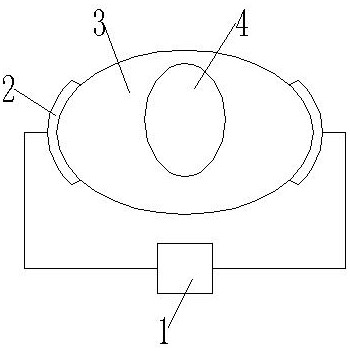
[0045] Such as Figure 1-5 As shown, a wearable bladder urine volume detection system includes: a parallel plate capacitor, a sensor, a single-chip microprocessor, a power module, an independent control key module and a display module, the parallel plate capacitor is connected to the sensor, and the sensor is connected to the The single-chip microprocessor is connected, and the single-chip microprocessor is connected with the power module, and the single-chip microprocessor is also connected with an independent button control module and a display module; the parallel plate capacitor includes two electrode sheets, one of which is the electrode One pad is set on one side of the bladder, and the other said electrode pad is set on the other side of the bladder. The sensor module includes a UTI03 sensor that monitors an analog signal of capacitance change. The independent control button module is used to control the working state of the system.
[0046] combine image 3As shown,...
Embodiment 2
[0052] A method for using a wearable bladder urine volume detection system includes the following steps:
[0053] S1: the installation of the parallel plate capacitor, the electrodes of the parallel plate capacitor are installed on both sides of the bladder;
[0054] S2: the measurement of capacitance value, measure the capacitance value of parallel plate capacitor by sensor, and the analog signal that monitors is transmitted to single-chip microprocessor;
[0055] S3: data processing, the single-chip microprocessor converts the received analog signal into a digital signal, and processes the data signal;
[0056] S4: the urine content is displayed, and the single-chip position processor displays the processed data through the display module;
[0057] S5: discharge of urine, when the urine content in the bladder reaches a preset threshold, the urine in the bladder is discharged.
Embodiment 3
[0059] combine Figure 8-10 As shown, a wearable device using a detection system includes a fixed belt 5, an elastic belt 6 is arranged in the middle of the fixed belt 5, and the elastic belt ensures the scalability of the fixed belt. The two ends of the fixed belt 5 Velcro 7 is provided, the two ends of the fixing band 5 are connected by Velcro 7, one side of the elastic band 6 is provided with an electrode sheet structure 8, the number of the electrode sheet structures 8 is two, and the two The electrode sheet structure 8 is arranged symmetrically about the elastic band 6, and the other side of the fixed band 5 is provided with a control button 14, a single-chip microprocessor 15 and a power module 16, and the control button is used to control the working state of the device. The power supply module 16 is used for providing electric energy to the single-chip microprocessor 15 .
[0060] A fixing groove 9 is arranged on the surface of the fixing belt 5 , and fixing pieces 10...
PUM
 Abstract
Description
Claims
Application Information
 - R&D
- Intellectual Property
- Life Sciences
- Materials
- Tech Scout
- Unparalleled Data Quality
- Higher Quality Content
- 60% Fewer Hallucinations
Browse by: Latest US Patents, China's latest patents, Technical Efficacy Thesaurus, Application Domain, Technology Topic, Popular Technical Reports.
© 2025 PatSnap. All rights reserved.Legal|Privacy policy|Modern Slavery Act Transparency Statement|Sitemap|About US| Contact US: help@patsnap.com


