Application of CEM in mammary duct angiography and verification method

A mammary duct, 1. CEM technology, applied in the field of application and verification of CEM in mammary duct angiography, can solve problems such as overlapping interference of mammary ducts, influence of ductal lesions, and influence on lesion diagnosis

Pending Publication Date: 2021-11-12
XIEHE HOSPITAL ATTACHED TO TONGJI MEDICAL COLLEGE HUAZHONG SCI & TECH UNIV
View PDF1 Cites 0 Cited by
  • Summary
  • Abstract
  • Description
  • Claims
  • Application Information

AI Technical Summary

Problems solved by technology

[0004] However, the conventional methods currently used have some limitations: the overlapping interference of mammary ducts affects the diagnosis of lesions; the display of ductal lesions in patients with dense breasts may be affected by the occlusion of their glands

Method used


Image

  • Application of CEM in mammary duct angiography and verification method
  • Application of CEM in mammary duct angiography and verification method
  • Application of CEM in mammary duct angiography and verification method

Examples


Embodiment 1

[0032] Step 1: Select an appropriate concentration of iodine-containing contrast agent, use a 1ml syringe combined with a No. 4 needle with a smooth tip to absorb the contrast agent, exhaust the air, and place it in a curved plate for later use. The concentration of iodine-containing contrast agent in this example is 300mg / ml.

[0033] Step 2: The patient is placed in a supine position, and the nipple on the affected side is disinfected according to the aseptic principle. The operator wears sterile gloves, and gently squeezes the vicinity of the areola and the nipple to promote nipple discharge, so as to find the correct milk duct opening.

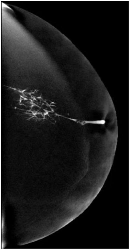
[0034] Step 3: Insert a ground No. 4 needle from the opening of the milk duct for about 0.5 to 1 cm. When injecting the drug, pinch the patient's nipple tightly to avoid overflow of the contrast agent. Slowly inject the contrast agent. When the patient's breast swelling is obvious, the needle is pulled out, and the angiography is performed...


PUM


Abstract

The invention discloses application of CEM in mammary duct angiography and a verification method. The verification method comprises the following steps of selecting an iodine-containing contrast agent with a proper concentration, and sucking the contrast agent for later use; enabling a patient to be in a supine position, wherein a nipple on the affected side is disinfected, and nipple discharge is promoted so that a correct breast duct opening can be found; injecting the contrast agent; and carrying out full-view digital mammography to obtain a delay period CEM image. The application of the CEM in mammary duct angiography is provided for the first time, it is verified through experiments that the CEM subtraction graph can eliminate the influence of compact glands around the duct on the focus, the size of the focus in the duct and branches at the far end of the duct can be better displayed, and the method has certain guiding significance in the clinical operation range.

Description

technical field [0001] The invention relates to the application of CEM, in particular to the application and verification method of CEM in mammary gland ductography. Background technique [0002] Contrast-enhanced mammography (CEM) uses the K-edge effect of iodine to quickly perform high- and low-energy dual-energy exposures after intravenous injection of contrast agents, and obtain low-energy images and subtraction images after post-processing. An emerging breast imaging technology. [0003] Nipple discharge is a relatively common symptom in breast diseases, and its main causes include tumor, ductal dilatation, inflammation, hyperplasia, etc., and mammary ductography is the main inspection method at present. Mammary duct radiography is a mammographic examination after injecting a contrast agent into the mammary duct, and according to the filling of the contrast agent, it can display the shape, expansion degree, location and distribution characteristics of the lesion in the...

Claims


Application Information

Patent Timeline
Patent Type & Authority Applications(China)
IPC IPC(8): A61B6/00
CPCA61B6/481A61B6/502
Inventor 杨帆陈乐庆陶娟朱文颖刘洁彭舒怡杨金荣
Owner XIEHE HOSPITAL ATTACHED TO TONGJI MEDICAL COLLEGE HUAZHONG SCI & TECH UNIV
Why Eureka
Try Eureka