Eye fundus retina image segmentation method based on deep convolutional neural network

An image segmentation and retinal technology, applied in the field of medical image processing, can solve problems such as inability to understand global information, loss of feature information, hinder segmentation performance, etc., and achieve the effect of improving extraction and recognition capabilities, maintaining relevance, and improving understanding capabilities

Pending Publication Date: 2021-12-07
NORTHWEST NORMAL UNIVERSITY
View PDF0 Cites 2 Cited by
  • Summary
  • Abstract
  • Description
  • Claims
  • Application Information

AI Technical Summary

Problems solved by technology

However, these methods have certain problems: 1) simple image preprocessing methods cannot effectively utilize the structural information of the feature map, which may hinder the segmentation performance; 2) use downsampling to improve the receptive field, but jointly segment the optic disc and cup , because the optic disc area on the label map is relatively small, an e

Method used


Image

  • Eye fundus retina image segmentation method based on deep convolutional neural network
  • Eye fundus retina image segmentation method based on deep convolutional neural network
  • Eye fundus retina image segmentation method based on deep convolutional neural network

Examples


Embodiment 1

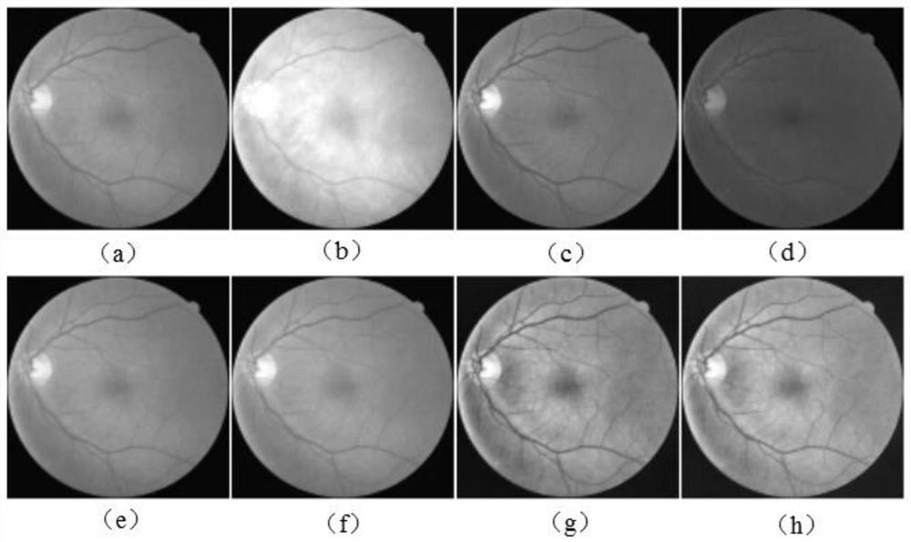
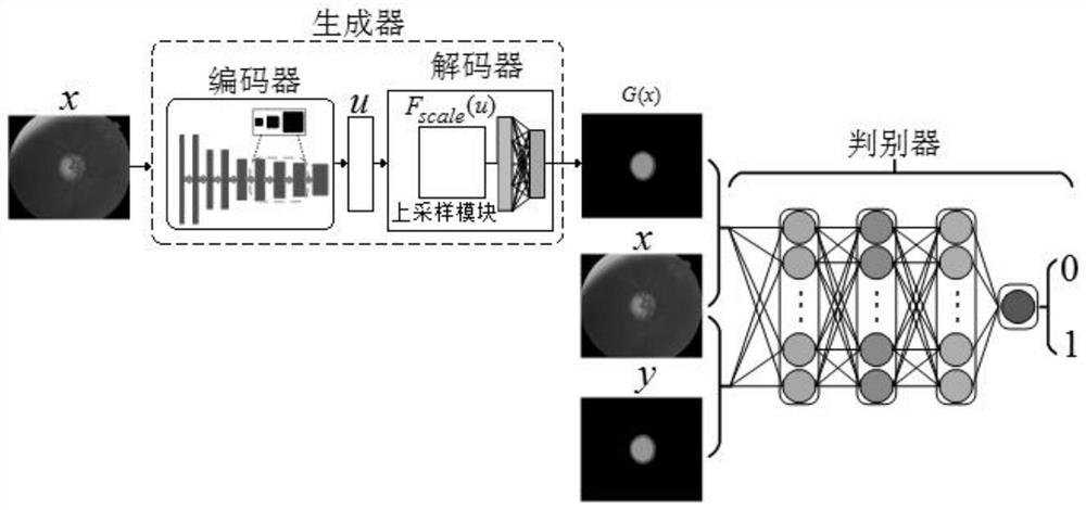
[0055] The segmentation method of the present invention is a method based on a deep convolutional neural network, and involves a data preprocessing model of a fundus retinal image, an end-to-end multi-label deep convolutional network model and the application of the model to the segmentation of retinal blood vessels, optic discs and optic cups.

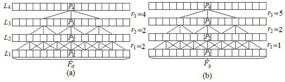
[0056] The segmentation method of the present invention uses a deep convolutional neural network to map the characteristics of blood vessel tissue, optic disc and optic cup tissue, and diseased tissue in medical images, and uses the convolutional network to segment the image. In addition, in order to increase the segmentation accuracy, a new data preprocessing method for fundus retinal images is used to enhance image processing; an end-to-end deep convolutional network is used to solve the problem of small blood vessel segmentation, and the deep salient features of the lesion area are obtained and visualized; using The method of combin...


PUM


Abstract

The invention discloses a fundus retina image segmentation method based on a deep convolutional neural network. The method employs the deep convolutional neural network to map features of a vascular tissue, an optic disc optic cup tissue and a lesion tissue in a medical image, and employs the convolutional network to segment the image. In addition, in order to increase segmentation accuracy, a new data preprocessing method of the fundus retina image is used for enhancing the image; an end-to-end deep convolutional network is used for solving a problem of small blood vessel segmentation, and deep significant features of a lesion area are obtained and visualized; a series of problems caused by large pixels of various medical images are solved by using a method of combining multiple deep neural networks.

Description

technical field [0001] The invention belongs to the technical field of medical image processing, and relates to a method for segmenting a retinal fundus image, in particular to a method for segmenting a fundus retinal image based on a deep convolutional neural network. Background technique [0002] Retinal images have been widely used in the diagnosis, screening, and treatment of cardiovascular and ophthalmic diseases, including systemic diseases such as age-related macular degeneration, glaucoma, hypertension, arteriosclerosis, and cardiovascular disease. Vessel, optic disc, and optic cup segmentation are fundamental steps required for quantitative analysis of retinal images. First, the division of the optic disc and optic cup is usually used for the detection of glaucoma. The optic nerve head assessment method is a convenient and widely accepted method used by clinicians. This method calculates the risk of glaucoma by using the ratio of the vertical cup-to-disk ratio, so ...

Claims


Application Information

Patent Timeline
IPC IPC(8): G06T5/40G06T7/00G06T7/11G06T7/90G06N3/04G06K9/62
CPCG06T5/40G06T7/11G06T7/90G06T7/0012G06T2207/30041G06N3/045G06F18/214
Inventor 蒋芸高静王发林姚慧霞马泽琪张婧瑶
Owner NORTHWEST NORMAL UNIVERSITY
Why Eureka
Try Eureka