
Chromosome division phase positioning and sorting method based on multi-scale feature fusion

A technology of multi-scale features and chromosomes, applied in neural learning methods, instruments, biological neural network models, etc., can solve the problem that it is difficult to achieve good results in detecting images, and achieve network degradation problems, reduce errors, and analyze speed Enhanced effect

Active Publication Date: 2021-12-17
SHANGHAI BEION MEDICAL TECH CO LTD
View PDF6 Cites 0 Cited by
  • Summary
  • Abstract
  • Description
  • Claims
  • Application Information

AI Technical Summary

Problems solved by technology

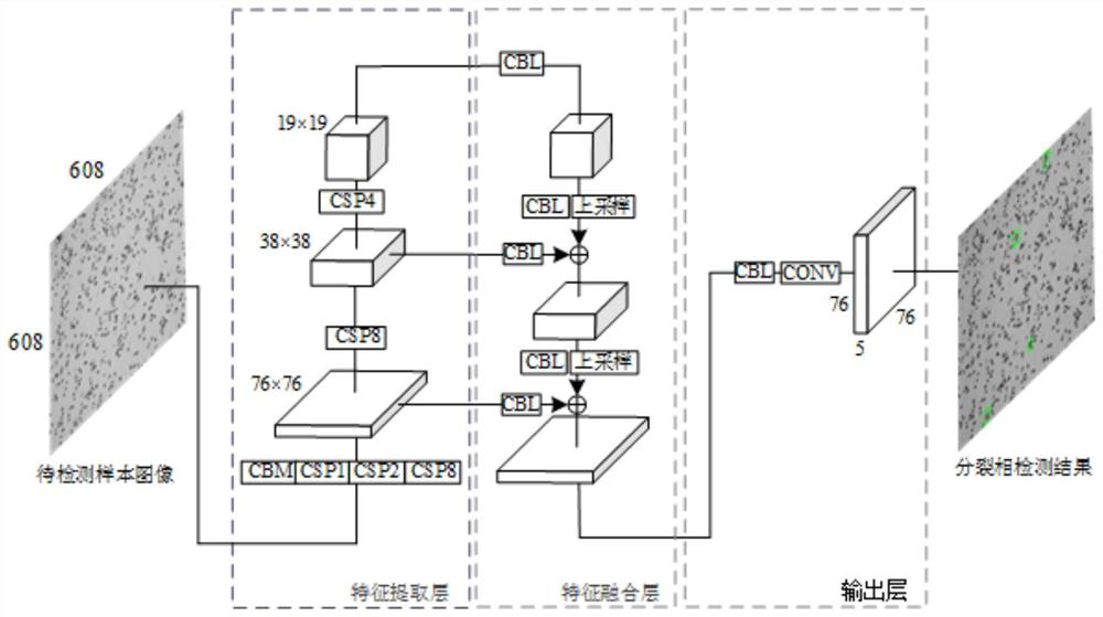
The current popular deep learning image analysis methods usually use simple linear networks or convolutional neural networks for modeling and analysis. These methods usually use multi-layer neural networks to extract abstract semantic features of images to complete image classification. Difficult to achieve good results with smaller targets

Method used


Image

  • Chromosome division phase positioning and sorting method based on multi-scale feature fusion
  • Chromosome division phase positioning and sorting method based on multi-scale feature fusion
  • Chromosome division phase positioning and sorting method based on multi-scale feature fusion

Examples


Embodiment Construction

[0062] The implementation of the present application will be described in detail below with reference to the accompanying drawings and examples, so as to fully understand and implement the implementation process of how the present application uses technical means to solve technical problems and achieve technical effects.

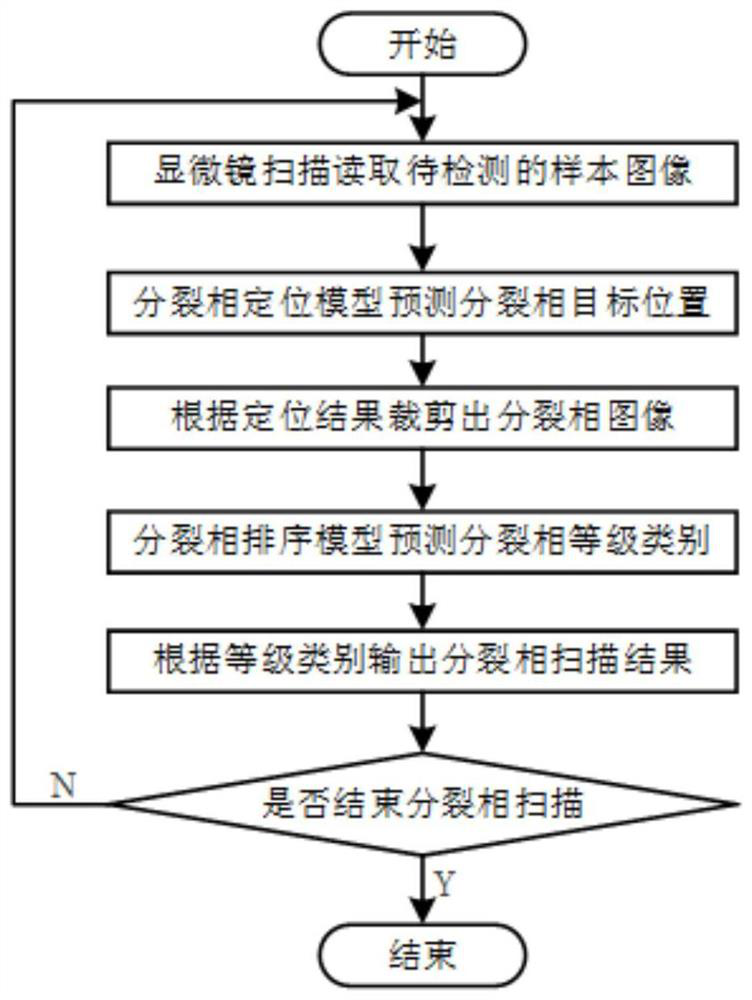
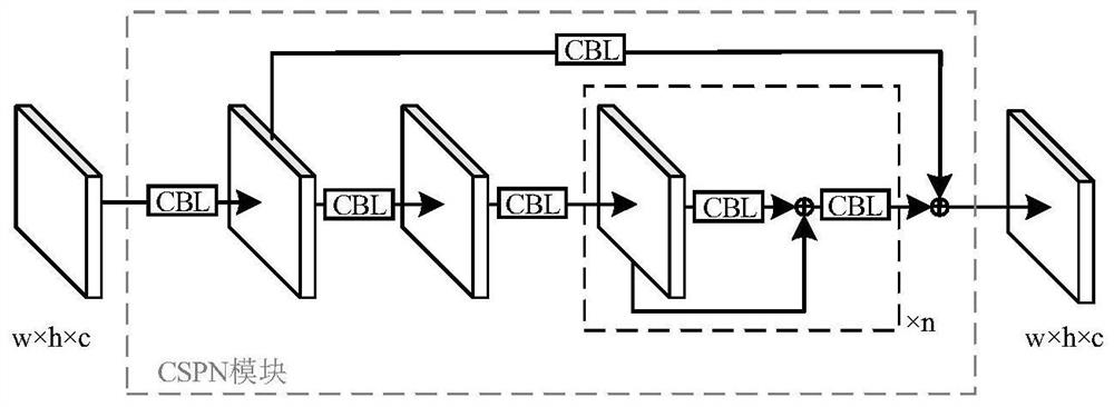
[0063] Compared with using traditional computer graphics and machine learning methods to locate chromosome cleavage phases, the deep learning method can use a large number of labeled samples for model training without relying on artificially extracted cleavage phase features. Under the premise that the scale is guaranteed, the model can often achieve better generalization. Based on deep learning technology, the present invention proposes a chromosome division phase location and sorting method based on multi-scale feature fusion. The method first uses convolutional neural network to extract the features of chromosome sample images on multiple scales, and then ...


PUM


Abstract

The invention discloses a chromosome division phase positioning and sorting method based on multi-scale feature fusion. The method comprises the following steps: S1, starting; S2, acquiring a training set; S3, obtaining an enhanced chromosome sample image; S4, acquiring an input tensor A; S5, training a split phase positioning model; S6, finally obtaining a training set for model training; S7, finally obtaining an input tensor B used for training the chromosome division phase sorting model; S8, outputting a split phase scanning result; and S9, judging whether split phase scanning is ended or not, if not, skipping to the step S2, and otherwise, ending. According to the method, feature extraction and sorting are carried out on the segmented split-phase images by training the deep learning model, so that the sorting effect can be greatly improved, parallel computing is carried out by splicing the segmented split-phase images into a tensor, and the influence of additional model calculation on the algorithm detection speed can be further reduced.

Description

technical field [0001] The invention belongs to a method for locating and sorting chromosome division phases based on multi-scale feature fusion. Background technique [0002] Chromosome karyotype analysis takes the mid-phase of chromosome division as the research object, uses banding technology and makes full use of the morphological and texture features of chromosomes to sort and number chromosomes, so as to complete the analysis of chromosomes, and karyotype analysis is cytogenetic analysis Research provides an important basis. The key to the automatic analysis of chromosome karyotypes using the microscope automatic scanning platform is to be able to use the computer to complete the location of chromosome cleavage phases and sort the quality of the extracted cleavage phases, so as to obtain more research-worthy cleavage phase positions. Then the cleavage phase image is segmented according to the specific position, so as to carry out the corresponding karyotype analysis. ...

Claims


Application Information

Patent Timeline
Patent Type & Authority Applications(China)
IPC IPC(8): G06K9/00G06K9/32G06K9/62G06N3/04G06N3/08
CPCG06N3/08G06N3/045G06F18/214
Inventor 崔玉峰许威
Owner SHANGHAI BEION MEDICAL TECH CO LTD
Learn More