Pulmonary nodule image detection method and system based on CT image

A CT image and image detection technology, applied in the field of image processing, can solve problems such as unsatisfactory results, and achieve the effect of avoiding subjective uncertainty and improving accuracy.

Pending Publication Date: 2022-01-04
WUHAN UNIV OF SCI & TECH
View PDF0 Cites 4 Cited by
  • Summary
  • Abstract
  • Description
  • Claims
  • Application Information

AI Technical Summary

Problems solved by technology

[0004] The present invention aims at the problem that the existing Transformer is only used to encode the marked image block, and then directly upsampling the hidden feature representation into a full-resolution dense output, which cannot produce satisfactory results. This type of architecture is usually used between patients. There are large differences in texture, shape and size. In order to overcome this limitation, the present invention provides a method and system for detecting pulmonary nodules based on CT images. The detection method uses the TransformerUnet combined architecture. On the basis of research, a self-attention mechanism based on CNN features (ie, self-attention mechanism) is proposed. Unlike existing CNN-based methods, TransformerUnet establishes a self-attention mechanism from the perspective of sequence-to-sequence prediction.

Method used


Image

  • Pulmonary nodule image detection method and system based on CT image
  • Pulmonary nodule image detection method and system based on CT image
  • Pulmonary nodule image detection method and system based on CT image

Examples


Embodiment 1

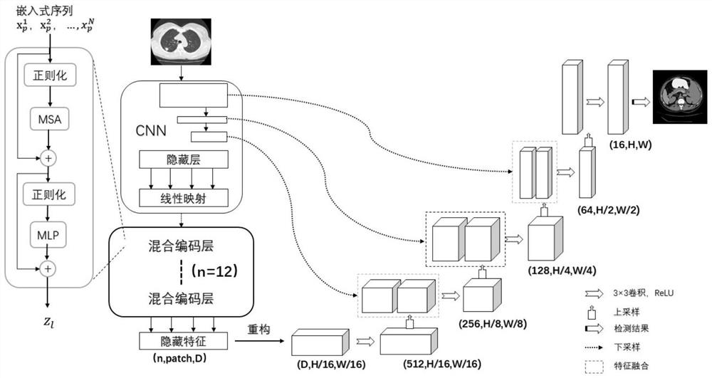
[0051] Such as figure 1 with figure 2 As shown, this embodiment discloses a CT image-based lung nodule image detection method, which uses a TransformerUnet combined architecture for lung nodule image detection, and the TransformerUnet combined architecture includes a Transformer part and a U-Net part.

[0052] The lung nodule image detection method based on CT image described in the present embodiment comprises steps as follows:

[0053] Step S1 , image preprocessing. The image preprocessing described in this embodiment adopts image serialization, that is, the slices of the input lung CT image are reshaped into a set of patch sequences to perform tokenization.

[0054] Let the given lung CT image be H×W is the spatial resolution, and C is the number of channels. The goal is to predict the corresponding pixel label map of size H×W, which is different from the existing training CNN (such as U-Net), which encodes the image into a high-level feature representation, and then d...

Embodiment 2

[0083] This embodiment discloses a system of a pulmonary nodule image detection method based on the CT image described in Embodiment 1, which adopts TransformerUnet combined architecture, specifically including:

[0084] An image serialization module for reshaping slices of input lung CT images into a set of patch sequences to perform tokenization;

[0085] A patch embedding module for mapping vectorized patch sequences to a latent two-dimensional embedding space using a trainable linear map;

[0086] A hybrid encoder module of CNN and Transformer, which is used to encode the tokenized image patches from the CNN feature map into the input sequence for extracting the global context through the Transformer;

[0087] The cascaded decoder module is used to first upsample the encoded features obtained by the hybrid encoder module of CNN and Transformer through the decoder, and then combine the upsampled encoded features with high-resolution CNN feature maps to achieve precise posit...


PUM


Abstract

The invention discloses a pulmonary nodule image detection method and system based on a CT image. The detection method comprises the following steps: S1, image serialization: remodeling slices of an input lung CT image into a group of patch sequences to execute marking; S2, utilizing patch embedding, and using trainable linear mapping to map the vectorized patch sequence to a potential two-dimensional embedding space; S3, establishing a hybrid encoder of the CNN and a Transformer: encoding the marked image blocks from the CNN feature map into an input sequence for extracting a global context through the Transformer; and S4, cascading a decoder: firstly performing up-sampling on the coding features obtained in the step S3 through the decoder, then combining the up-sampled coding features with a high-resolution CNN feature map to realize accurate positioning, and finally enhancing more accurate detail detection information by recovering local space information by using U-Net. According to the invention, the accuracy of pulmonary nodule detection can be effectively improved.

Description

technical field [0001] The present invention relates to the technical field of image processing, in particular to a method and system for detecting pulmonary nodules based on CT images. Background technique [0002] Lung cancer is the leading cause of death in the world. Pulmonary nodules, as an early manifestation of lung cancer, can be observed on CT images as round lung shadows with a diameter of no more than 3cm. Accurate detection of their contours can help doctors realize lung cancer. Diagnosis of benign and malignant nodules. Due to the small size of pulmonary nodules, their shape, brightness and other characteristics are similar to those of blood vessels and other tissues in the lung parenchyma. It is difficult to separate the two only by naked eye observation, which is likely to seriously interfere with the doctor's judgment. In order to reduce the workload of doctors and improve the efficiency of nodule diagnosis, computer-aided diagnosis technology has been used ...

Claims


Application Information

Patent Timeline
Patent Type & Authority Applications(China)
IPC IPC(8): G06T7/00G06N3/04G06N3/08
CPCG06T7/0012G06N3/08G06T2207/10081G06T2207/20081G06T2207/20084G06T2207/30064G06N3/045
Inventor 李波徐麒皓
Owner WUHAN UNIV OF SCI & TECH
Why Eureka
Try Eureka