Ultrasonic simulation method and system based on medical tomographic image
A technology of tomographic imaging and simulation method, applied in the field of ultrasound imaging, can solve the problems of unrealistic results, lack of tissues and organs, low resolution, etc., and achieve the effect of good geometric shape
- Summary
- Abstract
- Description
- Claims
- Application Information
AI Technical Summary
Problems solved by technology
Method used
Image
Examples
Embodiment 1
[0047] Embodiment 1 provides an ultrasound simulation system based on medical tomographic images, the system includes:
[0048] A calculation module, configured to calculate the acoustic impedance coefficient of each voxel of the tomographic image;
[0049] The first building block is used to perform reflection modeling on the tomographic image in combination with acoustic impedance coefficients to obtain an initial reflectivity map;
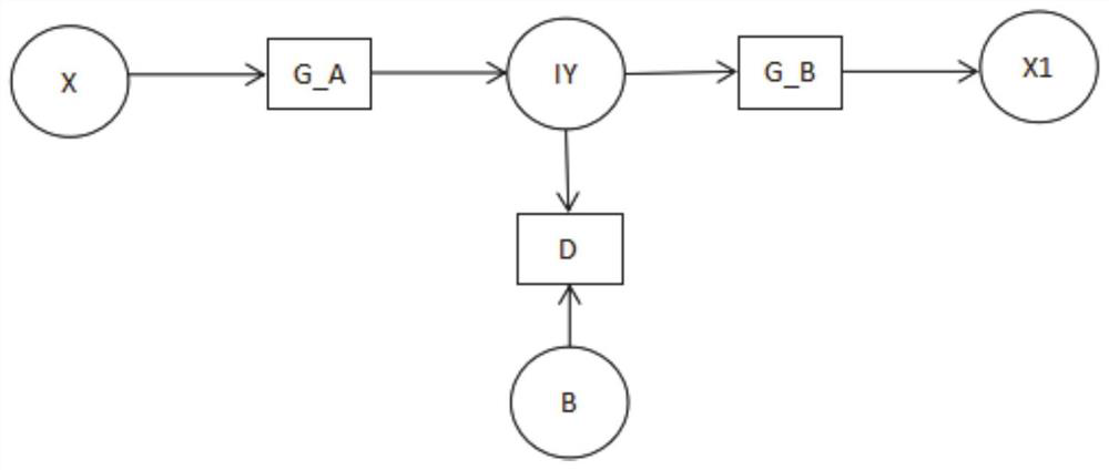
[0050] The second building block is used to process the initial reflectance map by using the reflectance generation network to obtain a complete reflectance map;
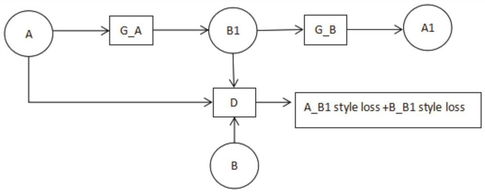
[0051] The third building block is used to simultaneously input the complete reflectance map and the real ultrasound image into the ultrasound deep learning network for realistic learning, and obtain the ultrasound virtual image.
[0052] In this embodiment 1, the ultrasound simulation method based on the medical tomographic image is realized by using the above-mentioned ultrasound s...
Embodiment 2
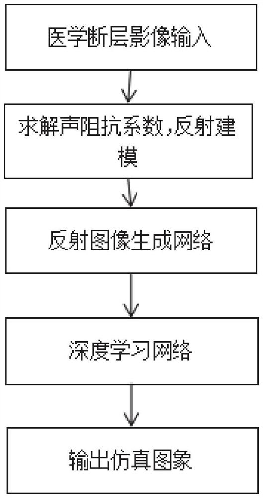
[0090] Such as figure 1 As shown, in this embodiment 2, an ultrasonic simulation method based on medical tomographic images is provided. First, the CT value is used to solve the acoustic impedance coefficient corresponding to each voxel by using the linear fitting method for the medical tomographic images; and then according to the obtained The acoustic impedance coefficient of the medical tomographic image is used for reflection modeling, and the method used is the traditional ray tracing algorithm; finally, the reflectance image generated by the modeling and the real ultrasound image data set are put into the deep learning network to generate the ultrasound simulation image . Include the following steps:
[0091] Step 1: Obtain the acoustic impedance coefficient of each voxel of the medical tomographic image;
[0092] Step 2: Use ray tracing algorithm to perform reflection modeling on the medical tomographic image injected with contrast agent;
[0093] Step 3: Use CUDA to...
Embodiment 3
[0136] Embodiment 3 of the present invention provides a non-transitory computer-readable storage medium. The non-transitory computer-readable storage medium is used to store computer instructions. When the computer instructions are executed by a processor, the above-mentioned medical-based An ultrasound simulation method for a tomographic image, the method comprising:
[0137] Calculate the acoustic impedance coefficient of each voxel of the tomographic image;
[0138] Combined with the acoustic impedance coefficient, the reflection modeling of the tomographic image is carried out to obtain the initial reflectivity map;
[0139] Use the albedo generation network to process the initial albedo map to obtain the complete albedo map;
[0140] The complete reflectance map and the real ultrasound image are simultaneously input into the ultrasound deep learning network for realistic learning, and the ultrasound virtual image is obtained.
PUM
 Abstract
Description
Claims
Application Information
 - R&D
- Intellectual Property
- Life Sciences
- Materials
- Tech Scout
- Unparalleled Data Quality
- Higher Quality Content
- 60% Fewer Hallucinations
Browse by: Latest US Patents, China's latest patents, Technical Efficacy Thesaurus, Application Domain, Technology Topic, Popular Technical Reports.
© 2025 PatSnap. All rights reserved.Legal|Privacy policy|Modern Slavery Act Transparency Statement|Sitemap|About US| Contact US: help@patsnap.com


