Animal eyeball pathological section manufacturing method

A technology of pathological slices and production methods, applied in the field of life sciences, can solve the problems of inability to form high-precision slices, easy deformation of eyeball tissue, thick pathological slices, etc., and achieve efficient and sufficient dehydration treatment, excellent observability, and complete structure.

Active Publication Date: 2022-02-25
WEST CHINA HOSPITAL SICHUAN UNIV
View PDF10 Cites 2 Cited by
  • Summary
  • Abstract
  • Description
  • Claims
  • Application Information

AI Technical Summary

Problems solved by technology

[0008] The purpose of the present invention is to provide a method for making pathological slices of animal eyeballs in view of the problems existing in the prior art that the eyeball tissue is easily deformed and lost, the pathological slices are thick and cannot form continuous high-precision slices

Method used


Image

  • Animal eyeball pathological section manufacturing method
  • Animal eyeball pathological section manufacturing method
  • Animal eyeball pathological section manufacturing method

Examples


Embodiment 1

[0063] Example 1 Production of pathological slices of animal eyeballs

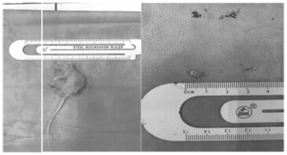
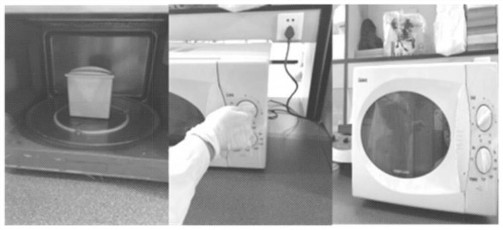
[0064] Such as Figure 1-Figure 7 As shown, mouse eye pathological sections were made. The eyeballs of the animals were removed and the 6mm optic nerve was reserved, and the blood stains on the surface of the eyeballs were removed by washing with normal saline. Then, the removed animal eyeballs were placed in physiological saline, and then heated in a microwave oven for 5 minutes.

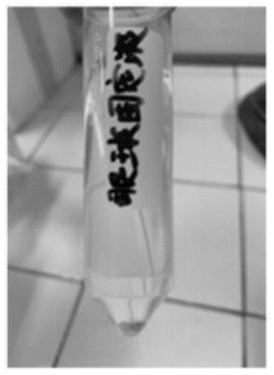
[0065] Take 20ml of glacial acetic acid, 60ml of chloroform, and 120ml of methanol, and mix them to make a fixative. The eyeballs were placed in 50mL fixative solution and fixed at room temperature 18-22°C for 72h. Take out the mouse eyeball from the fixative solution, wash it with running water for 2 hours, put the eyeball tissue in a wide-mouth bottle when washing, tie the bottle mask with gauze and fasten it with a thread, put it under the tap with a rubber tube, and insert the water outlet end of the rubber tube into t...

Embodiment 2-7

[0074] Example 2-7 Production of pathological slices of animal eyeballs

[0075] Animal eyeball pathology sections were prepared in the same way as in Example 1, the only difference being that after the animal eyeball was removed, the intensity of heating treatment with medium heat in a microwave oven was different. In this example, microwaves were heated for 4-10 minutes on low, medium, and high heat, and then processed according to the same process method as in Example 1, and animal eyeball slices were finally prepared. The results are shown in the table below.

[0076] Table 1 Effect of microwave treatment intensity on morphology of animal eyeball slices

[0077] Example Firepower processing time Processing effect slice integrity 2 medium fire 4min good shape whole 3 medium fire 10min good shape whole 4 small fire 4min General shape small defect 5 small fire 10min General shape small defect 6 fire 4...

Embodiment 8-11

[0080] Examples 8-11 Production of pathological slices of animal eyeballs

[0081] The eyeballs of the animals were removed and the 6mm optic nerve was reserved, and the blood stains were removed by flushing with normal saline. Then, the removed animal eyeballs were placed in physiological saline, and then heated in a microwave oven for 5 minutes.

[0082]Take 20ml of glacial acetic acid, 60ml of chloroform, and 120ml of methanol, and mix them to make a fixative. The eyeballs were placed in 50mL of fixative solution and fixed at room temperature for 72h. The eyeballs of the mice were taken out from the fixative solution, rinsed with running water for 2 hours, and the tissue was placed in a jar when washing, and the bottle was covered with gauze and fastened with a thread, and the eyeball was confined in the jar with gauze. Place the wide-mouth bottle under the tap with a rubber tube inserted into the bottle, allowing the water to slowly overflow from the bottom of the bott...


PUM

PropertyMeasurementUnit
thicknessaaaaaaaaaa

Abstract

The invention relates to an animal eyeball pathological section manufacturing method which comprises the following steps: picking an animal eyeball, reserving 3-8mm optic nerve, and washing with normal saline to remove bloodstain; placing the animal eyeballs in normal saline, and then subjecting the animal eyeballs to heating treatment for 4-12 min with medium fire in a microwave oven; putting animal eyeballs into a stationary liquid prepared from glacial acetic acid, chloroform and methanol, and fixing for 60-90 hours; dehydrating the animal eyeballs 2-4 times by using n-butyl alcohol, wherein each time lasts for 0.5-3 hours; then, subjecting the animal eyeballs to transparent treatment with dimethylbenzene for 2-4 times, wherein each time lasts for 10-20 min, and the total time of dimethylbenzene transparent treatment is 30-50 min; performing the first paraffin treatment for 1-2 hours, performing the second paraffin treatment for 1-2 hours, and performing paraffin embedding after the treatment; and slicing according to a set thickness. When the animal eyeball pathological section is manufactured by the method, the eyeballs are treated by microwaves in combination with the structural characteristics of the animal eyeballs, so that the eyeball structural protein is denatured, the strength of the eyeball structural strength is pre-enhanced, and the method has important significance on accurate research and teaching of fine structures of the animal eyeballs.

Description

technical field [0001] The invention relates to a method for making animal specimens, in particular to a method for making animal eyeball pathological slices, and belongs to the technical field of life sciences. Background technique [0002] Animal specimens refer to special scientific research or teaching tools that are artificially processed to display the microscopic appearance of specific animal tissues. The existing technology has relatively rich experience in making common animal pathological slices, and the overall production process has also formed some relatively standardized operating procedures. However, for some special animal tissues or organs, due to other morphological and structural characteristics, the general specimen preparation The method is difficult to meet the production requirements of corresponding tissues or organs, and it is impossible to obtain satisfactory animal slice specimen samples. [0003] When dehydrating tissues or organs, due to the com...

Claims


Application Information

Patent Timeline
Patent Type & Authority Applications(China)
IPC IPC(8): G01N1/28
CPCG01N1/2813
Inventor 王竹陈洁田昆韩佳杞李稳王程仕谢钰鑫苟启桁黄芹锁娇娇胡靖睿李昀声
Owner WEST CHINA HOSPITAL SICHUAN UNIV
Why Eureka
Try Eureka