Biomarker for diagnosing abortion and application thereof
A marker and biological technology, applied in the field of biomarkers for diagnosing abortion, can solve the problems of limited evaluation value and limited accuracy of clinical diagnosis and prognosis of threatened abortion, and achieve the effect of promoting self-renewal ability.
- Summary
- Abstract
- Description
- Claims
- Application Information
AI Technical Summary
Problems solved by technology
Method used
Image
Examples
Embodiment 1
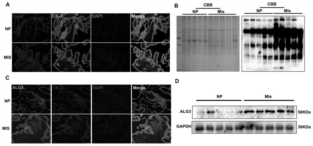
[0023] Clinical samples (villous tissue) from patients with inevitable abortion and healthy women with normal pregnancy, embedded in paraffin.
[0024] The expressions of Manα1, 3Man and ALG3 in villi tissue were detected.
[0025] 1. Immunohistochemical detection of the localization and expression of Manα1,3Man and ALG3 in villi tissue
[0026] (1) Dewaxing: put the prepared paraffin sections into the following solutions in turn: soak in xylene I for 10 minutes, soak in xylene II for 10 minutes, soak in 100% ethanol I for 5 minutes, soak in 100% ethanol II for 5 minutes, soak in 95% ethanol for 5 minutes , soak in 85% ethanol for 5 minutes, soak in 70% ethanol for 5 minutes; then rinse with slow water for 10 minutes. Dip in PBS 3 times, 5min each time.
[0027] (2) Antigen retrieval: Put the slices into citrate buffer and thaw in a microwave oven for 20 minutes. Take it out and let it cool down to room temperature naturally.
[0028] (3) Rinse with PBS 3 times, 5 min each...
Embodiment 2
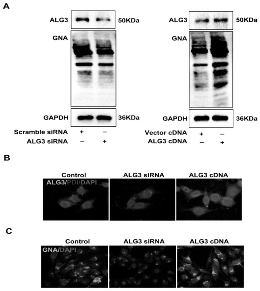
[0047] Example 2: Detection of Manα1,3Man expression in trophoblast cells by Lectin staining and cell immunofluorescence
[0048] Slip-up: After the trophoblast cells were digested with trypsin, centrifuge at 800 rpm for 4 minutes, resuspend in fresh medium, and put the cell suspension into a petri dish with slides.
[0049] Collection: remove the culture medium in the culture dish, wash with PBS 3 times, 3 minutes each time.
[0050] Fixation: add 4% paraformaldehyde to the petri dish and fix it for 20min.
[0051] Paraformaldehyde was discarded, washed 3 times with PBS, 3 min each time.
[0052] Blocking: block with immunostaining blocking solution for 1h.
[0053] Primary antibody incubation: overnight at 4°C (rabbit anti-human ALG3 1:150; lectin GNA 1:300). Wash with PBS 6 times, 5min each time.
[0054] Fluorescently labeled secondary antibodies were added dropwise, incubated at room temperature for 1 h, and washed 6 times with PBS for 5 min each time.
[0055] DAPI ...
Embodiment 3
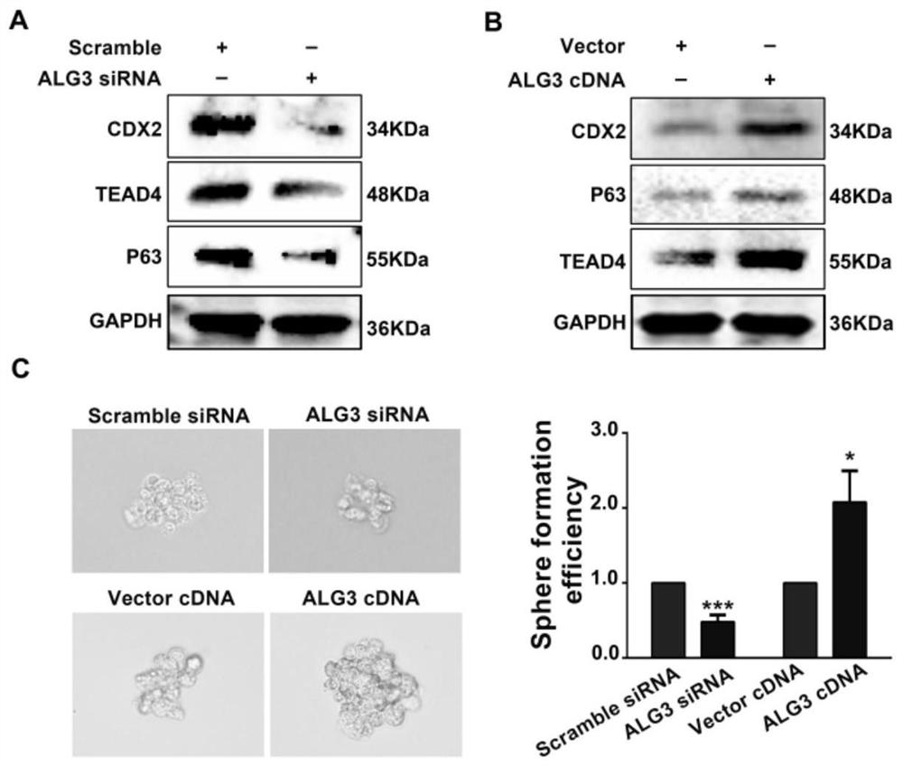
[0058] Example 3: Spheroid formation test to detect the self-renewal ability of trophoblast cells
[0059] (1) The trophoblast cells in good growth state were digested and centrifuged, the medium containing serum was removed, and washed twice with PBS.
[0060] (2) Prepare stem cell medium to resuspend cells (10 4 cells): DMEM / F12+1ⅹB27+20ng / mL bFGF+20ng / mL EGF.
[0061] (3) Cell plating: Choose a low-adhesion 6-well plate, with 4 ml of medium per well. The cultivation was completed in about 10 days, and the state of ball formation was observed.
[0062] Depend on image 3 It can be seen that transfection of ALG3 siRNA can inhibit the self-renewal ability of trophoblast cells and the stemness of trophoblast cells; while ALG3 cDNA can promote the self-renewal ability of trophoblast cells and the stemness of trophoblast cells.
PUM
 Abstract
Description
Claims
Application Information
 - R&D
- Intellectual Property
- Life Sciences
- Materials
- Tech Scout
- Unparalleled Data Quality
- Higher Quality Content
- 60% Fewer Hallucinations
Browse by: Latest US Patents, China's latest patents, Technical Efficacy Thesaurus, Application Domain, Technology Topic, Popular Technical Reports.
© 2025 PatSnap. All rights reserved.Legal|Privacy policy|Modern Slavery Act Transparency Statement|Sitemap|About US| Contact US: help@patsnap.com


