Observation device for clinical vaginal dilation internal penetration in obstetrics and gynecology department

An observation device, obstetrics and gynecology technology, applied in the direction of endoscopy, colposcopy, medical science, etc., can solve the problems of pollution, vaginal observation, patient discomfort, etc., and achieve the effect of convenient use, simple structure, and reduced discomfort

Inactive Publication Date: 2022-03-22
NANYANG SECOND GENERAL HOSPITAL
View PDF0 Cites 0 Cited by
  • Summary
  • Abstract
  • Description
  • Claims
  • Application Information

AI Technical Summary

Problems solved by technology

[0003] Now when performing gynecological examination, it is necessary to use a vaginal dilator, but the existing vaginal dilator has no light source or the light source is at the front end of the vaginal dilator, and can only illuminate the front part, making it difficult to observe the inside of the vagina comprehensively and carefully; After the vaginal dilator is stretched out, due to the cover of the support plate, it is impossible to observe the supported part. Some vaginal dilators have through holes for observation, but because there is no light source on the side, it is difficult to see the condition of the inner wall; the inspection process requires more When using the vaginal speculum for the first time, the vaginal speculum can only be placed on the table after taking it out, which may cause pollution. Moreover, when the existing vaginal speculum is used, especially in winter, the large temperature difference will bring strong stimulation to the patient feeling, making the patient uncomfortable

Method used


Image

  • Observation device for clinical vaginal dilation internal penetration in obstetrics and gynecology department
  • Observation device for clinical vaginal dilation internal penetration in obstetrics and gynecology department

Examples


Embodiment 1

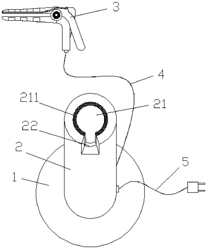
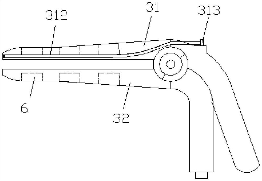
[0020] Embodiment one: Figure 1-Figure 2 It shows an obstetrics and gynecology clinical dilating and penetrating observation device, which is mainly composed of: a base 1, a placement cylinder 2, a dilating device 3, a connecting wire 4 and a power cord 5 Formed, the placement cylinder 2 is arranged on the base 1, the vaginal dilator 3 is connected with the placement cylinder 2 through the connecting wire 4, and the power cord 5 is arranged on the placement cylinder 2; The placement cylinder 2 is provided with a placement groove 21 for placing the vaginal expander; the inner wall of the placement groove 21 is provided with a heating layer 211 to heat the vaginal expander; the top of the placement cylinder 2 is provided with a card slot 22 , the venereal expander is in an "L" shape as a whole, and the card slot makes it more stable to place; the venereal expander 3 includes: an upper support plate 31 and a lower support plate 32, and the upper support plate 31 and the lower su...


PUM


Abstract

The invention discloses a clinical vaginal dilation inward entry observation device for the obstetrics and gynecology department, the clinical vaginal dilation inward entry observation device for the obstetrics and gynecology department is mainly formed by combining a base, a placing cylinder, a vaginal dilator, a connecting line and a power line, the placing cylinder is arranged on the base, the vaginal dilator is connected with the placing cylinder through the connecting line, and the power line is connected with the placing cylinder through the connecting line. The power line is arranged on the placing cylinder; the clinical vaginal dilation insertion observation device for the obstetrics and gynecology department is simple in structure and convenient to use, the whole device is transparent through the light guide optical fiber arranged in the upper supporting plate, the front end of a vaginal dilator and the side wall of the supported part can be observed through the opening, the vaginal dilator can be placed in the containing cylinder when taken down temporarily or before being used, and the clinical vaginal dilation insertion observation device for the obstetrics and gynecology department is clean, sanitary and convenient to use. And the vaginal dilator can be heated, so that the temperature is close to the body temperature of a human body, and the discomfort brought to a patient when the patient enters the vaginal dilator is reduced.

Description

technical field [0001] The invention relates to the field of medical auxiliary devices, in particular to an obstetrics and gynecology clinical dilation and penetrating observation device. Background technique [0002] Obstetrics and Gynecology is one of the four main disciplines of clinical medicine. It mainly studies the etiology, pathology, diagnosis and prevention of female reproductive organ diseases, the physiological and pathological changes of pregnancy and childbirth, the prevention, diagnosis and treatment of high-risk pregnancy and dystocia, female reproductive endocrinology, Family planning and women's health care. [0003] Now when performing gynecological examination, it is necessary to use a vaginal dilator, but the existing vaginal dilator has no light source or the light source is at the front end of the vaginal dilator, and can only illuminate the front part, making it difficult to observe the inside of the vagina comprehensively and carefully; After the va...

Claims


Application Information

Patent Timeline
Patent Type & Authority Applications(China)
IPC IPC(8): A61B1/32A61B1/303A61B1/07
CPCA61B1/32A61B1/303A61B1/07A61B1/0684
Inventor 谢志平
Owner NANYANG SECOND GENERAL HOSPITAL
Why Eureka
Try Eureka