
MRI (Magnetic Resonance Imaging) segmentation method for integrating attention mechanism aiming at brain lesion

A technology of attention and brain, applied in the field of image recognition, can solve the problems of limited receptive field of convolution kernel, high labeling requirements, and poor effect, so as to avoid calculation and memory requirements, improve lesion segmentation accuracy, increase Effect of color information data

Pending Publication Date: 2022-04-12
FUZHOU UNIV
View PDF0 Cites 6 Cited by
  • Summary
  • Abstract
  • Description
  • Claims
  • Application Information

AI Technical Summary

Problems solved by technology

[0005] The currently commonly used methods, in the face of small, long-distance and irregular white matter lesions, will cause segmentation errors; the current commonly used segmentation methods for brain MRI tissues and lesions usually use the neural network architecture, but in brain MRI The distribution of lesions is not the same even in different cases of the same data set. Only using these convolutional networks will make it difficult to establish a clear long-distance dependency due to the limited receptive field of the convolution kernel, so the lesions cannot be extracted well. The context information of the region leads to the final segmentation of brain lesions. At the same time, medical images usually have high requirements for labeling, the data set is small, and the effect of direct training is often not very good.

Method used


Image

  • MRI (Magnetic Resonance Imaging) segmentation method for integrating attention mechanism aiming at brain lesion
  • MRI (Magnetic Resonance Imaging) segmentation method for integrating attention mechanism aiming at brain lesion
  • MRI (Magnetic Resonance Imaging) segmentation method for integrating attention mechanism aiming at brain lesion

Examples


Embodiment Construction

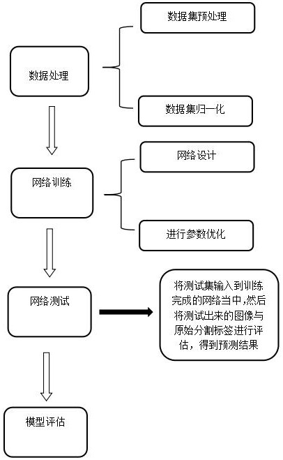
[0060] As shown in the figure, an MRI segmentation method for integrating brain lesions into the attention mechanism includes the following steps;

[0061] Step S1, collecting brain MRI images with segmentation map results, and establishing a training set;

[0062] Step S2, preprocessing the original brain MRI images to be segmented in the training set;

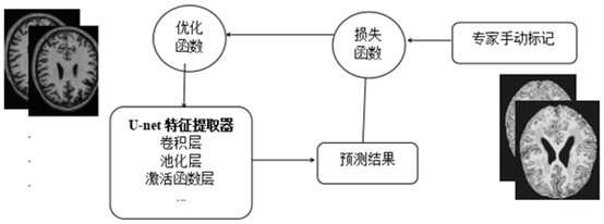
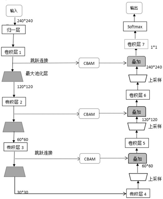
[0063] Step S3, establishing a convolutional neural network with an attention mechanism, and training its model with a training set;

[0064] Step S4. After the model training is completed, use the trained model parameters to predict the verification set images, and generate brain MRI tissue and lesion segmentation maps;

[0065] Step S5, establishing an evaluation file, and evaluating the segmentation result.

[0066] In step S1, use the MRBRains18 data set containing normal brain tissue and diseased tissue, and the MRI images of the MRBRains18 data set include three modes of T1, T2 and Flair; in step S1, perform skull str...


PUM


Abstract

The invention provides an MRI (Magnetic Resonance Imaging) segmentation method for integrating an attention mechanism aiming at brain lesions. The method comprises the following steps: S1, collecting a brain MRI image with a segmentation image result, and establishing a training set; s2, preprocessing the original brain MRI image to be segmented in the training set; s3, establishing a convolutional neural network with an attention mechanism, and training a model of the convolutional neural network by using the training set; s4, after model training is completed, using trained model parameters to predict the verification set image, and generating a brain MRI tissue and lesion segmentation map; s5, establishing an evaluation file, and evaluating a segmentation result; according to the method, more critical and important information can be extracted, and meanwhile, the training effect of training on a small data set is improved by means of transfer learning.

Description

technical field [0001] The invention relates to the technical field of image recognition, in particular to an MRI segmentation method for brain lesions integrated into an attention mechanism. Background technique [0002] Image segmentation has always been a research hotspot in the field of computer vision. Image segmentation is the technology and process of dividing an image into several specific regions with unique properties and proposing objects of interest. It is a key step from image processing to image analysis [0003] Magnetic resonance imaging (MRI) is a noninvasive medical imaging technique widely used in clinical diagnosis and brain research. It features high spatial resolution and fast acquisition time. Different MRI modalities, such as T1-weighted (T1), T2-weighted (T2), T1-weighted contrast-enhanced (T1C), and T2-weighted fluid-attenuated inversion recovery (FLAIR), provide different contrast information that can help distinguish between different types of ...

Claims


Application Information

Patent Timeline
Patent Type & Authority Applications(China)
IPC IPC(8): G06V10/26G06V10/44G06V10/774G06V10/82G06N3/04G06N3/08G06K9/62
Inventor 黄立勤庄炜杰
Owner FUZHOU UNIV
Learn More