Brain image processing method and system for stroke patient

An image processing and stroke technology, applied in the field of medical image processing, can solve the problems of obvious individual differences of stroke patients, excessive imaging area error, small brain images, etc., and achieve clear and accurate brain structure outline and lesion characteristics. Solve the problem of error recognition and improve the effect of recognition accuracy

Active Publication Date: 2022-04-15
FOSHAN UNIVERSITY +1
View PDF3 Cites 2 Cited by
  • Summary
  • Abstract
  • Description
  • Claims
  • Application Information

AI Technical Summary

Problems solved by technology

However, these two methods only use pixel-level features for image processing, and ignore the structural features of the brain when they are applied to brain image processing, which will lead to a certain degree of limitation in the accuracy of brain contour recognition.
However, the extraction of brain image features based on deep learning algorithms requires a large number of training set data samples, and the individual differences among stroke patients are obvious. The available medical image data cannot satisfy a large number of data samples, which is likely to cause overfitting problems. training results
In addition, due to the variability of brain atrophy or local vascular rupture in stroke patients, the brain image is small and there are many shadow areas. risk of misdiagnosis

Method used


Image

  • Brain image processing method and system for stroke patient
  • Brain image processing method and system for stroke patient
  • Brain image processing method and system for stroke patient

Examples


Embodiment Construction

[0076] The concept, specific structure and technical effects of the present disclosure will be clearly and completely described below in conjunction with the embodiments and drawings, so as to fully understand the purpose, scheme and effect of the present disclosure. It should be noted that, in the case of no conflict, the embodiments in the present application and the features in the embodiments can be combined with each other.

[0077] In the description of the present invention, the diagrams provided in the following embodiments are only schematically illustrating the basic ideas of the present invention, and only the components related to the present invention are shown in the diagrams rather than the number of components in actual implementation. , shape and village drawing, the type, quantity and proportion of each component can be changed arbitrarily during its actual implementation, and its component layout type may also be more complicated.

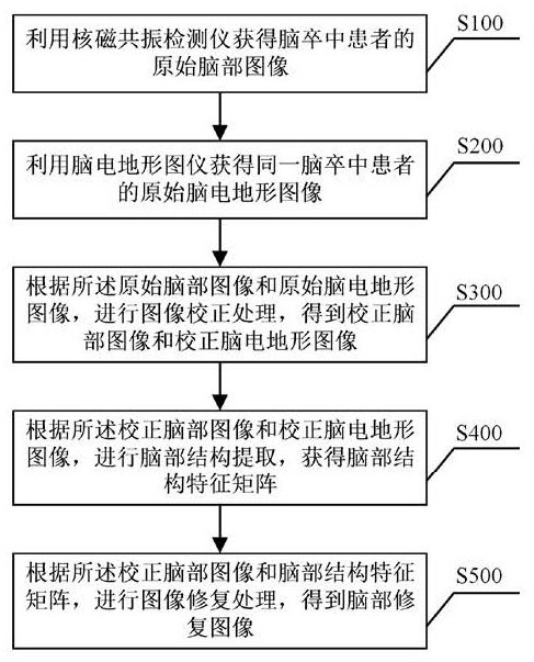
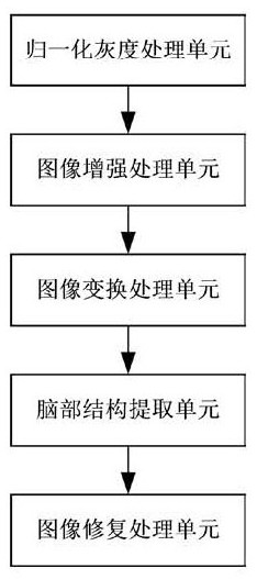
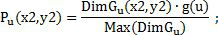
[0078] Such as figure 1 ...


PUM


Abstract

The invention discloses a brain image processing method and system for a stroke patient, and the method comprises the steps: obtaining an original brain structure image and an electroencephalogram topographic image of the stroke patient through a nuclear magnetic resonance detector and an electroencephalogram topographic map instrument; and data conversion preprocessing, image conversion processing, brain structure extraction, image restoration processing and the like are carried out, so that a brain restoration image with relatively high recognition accuracy is obtained. According to the brain image processing method and system, the shadow blurred area in the original brain structure image can be corrected, so that the brain structure contour is clearer and more accurate, and the recognition accuracy of the brain structure contour in the MRI brain image is improved.

Description

technical field [0001] The invention relates to the field of medical image processing, in particular to a brain image processing method and system for stroke patients. Background technique [0002] Stroke is an acute cerebrovascular disease with high morbidity and mortality. Mainly including ischemic stroke and hemorrhagic stroke, the pathogenic mechanism is brain tissue damage due to rupture or blockage of local cerebral blood vessels. With the help of medical imaging technology, real artifact images of brain tissue of stroke patients can be obtained, which can effectively provide more accurate physiological information to help doctors make early clinical diagnosis. Currently, clinical brain imaging techniques such as computed tomography (CT), positron emission tomography (PET), magnetic resonance imaging (Magnetic Resonance Imaging, MRI) are widely used. Because MRI can obtain high spatial resolution brain anatomical structure, and does not need to inject radioactive sub...

Claims


Application Information

Patent Timeline
IPC IPC(8): G06T7/00G06N20/00G06T7/12G06T7/136G06T7/66
CPCG06T7/0004G06T7/12G06T7/136G06T7/66G06N20/00G06T2207/10088G06T2207/20081G06T2207/30016
Inventor 孙金燕庞日宠郝志峰杨安平谭海曙艾毅龙赵超超
Owner FOSHAN UNIVERSITY
Why Eureka
Try Eureka